2.24: Cell Division
- Page ID
- 8921
\( \newcommand{\vecs}[1]{\overset { \scriptstyle \rightharpoonup} {\mathbf{#1}} } \)
\( \newcommand{\vecd}[1]{\overset{-\!-\!\rightharpoonup}{\vphantom{a}\smash {#1}}} \)
\( \newcommand{\dsum}{\displaystyle\sum\limits} \)
\( \newcommand{\dint}{\displaystyle\int\limits} \)
\( \newcommand{\dlim}{\displaystyle\lim\limits} \)
\( \newcommand{\id}{\mathrm{id}}\) \( \newcommand{\Span}{\mathrm{span}}\)
( \newcommand{\kernel}{\mathrm{null}\,}\) \( \newcommand{\range}{\mathrm{range}\,}\)
\( \newcommand{\RealPart}{\mathrm{Re}}\) \( \newcommand{\ImaginaryPart}{\mathrm{Im}}\)
\( \newcommand{\Argument}{\mathrm{Arg}}\) \( \newcommand{\norm}[1]{\| #1 \|}\)
\( \newcommand{\inner}[2]{\langle #1, #2 \rangle}\)
\( \newcommand{\Span}{\mathrm{span}}\)
\( \newcommand{\id}{\mathrm{id}}\)
\( \newcommand{\Span}{\mathrm{span}}\)
\( \newcommand{\kernel}{\mathrm{null}\,}\)
\( \newcommand{\range}{\mathrm{range}\,}\)
\( \newcommand{\RealPart}{\mathrm{Re}}\)
\( \newcommand{\ImaginaryPart}{\mathrm{Im}}\)
\( \newcommand{\Argument}{\mathrm{Arg}}\)
\( \newcommand{\norm}[1]{\| #1 \|}\)
\( \newcommand{\inner}[2]{\langle #1, #2 \rangle}\)
\( \newcommand{\Span}{\mathrm{span}}\) \( \newcommand{\AA}{\unicode[.8,0]{x212B}}\)
\( \newcommand{\vectorA}[1]{\vec{#1}} % arrow\)
\( \newcommand{\vectorAt}[1]{\vec{\text{#1}}} % arrow\)
\( \newcommand{\vectorB}[1]{\overset { \scriptstyle \rightharpoonup} {\mathbf{#1}} } \)
\( \newcommand{\vectorC}[1]{\textbf{#1}} \)
\( \newcommand{\vectorD}[1]{\overrightarrow{#1}} \)
\( \newcommand{\vectorDt}[1]{\overrightarrow{\text{#1}}} \)
\( \newcommand{\vectE}[1]{\overset{-\!-\!\rightharpoonup}{\vphantom{a}\smash{\mathbf {#1}}}} \)
\( \newcommand{\vecs}[1]{\overset { \scriptstyle \rightharpoonup} {\mathbf{#1}} } \)
\(\newcommand{\longvect}{\overrightarrow}\)
\( \newcommand{\vecd}[1]{\overset{-\!-\!\rightharpoonup}{\vphantom{a}\smash {#1}}} \)
\(\newcommand{\avec}{\mathbf a}\) \(\newcommand{\bvec}{\mathbf b}\) \(\newcommand{\cvec}{\mathbf c}\) \(\newcommand{\dvec}{\mathbf d}\) \(\newcommand{\dtil}{\widetilde{\mathbf d}}\) \(\newcommand{\evec}{\mathbf e}\) \(\newcommand{\fvec}{\mathbf f}\) \(\newcommand{\nvec}{\mathbf n}\) \(\newcommand{\pvec}{\mathbf p}\) \(\newcommand{\qvec}{\mathbf q}\) \(\newcommand{\svec}{\mathbf s}\) \(\newcommand{\tvec}{\mathbf t}\) \(\newcommand{\uvec}{\mathbf u}\) \(\newcommand{\vvec}{\mathbf v}\) \(\newcommand{\wvec}{\mathbf w}\) \(\newcommand{\xvec}{\mathbf x}\) \(\newcommand{\yvec}{\mathbf y}\) \(\newcommand{\zvec}{\mathbf z}\) \(\newcommand{\rvec}{\mathbf r}\) \(\newcommand{\mvec}{\mathbf m}\) \(\newcommand{\zerovec}{\mathbf 0}\) \(\newcommand{\onevec}{\mathbf 1}\) \(\newcommand{\real}{\mathbb R}\) \(\newcommand{\twovec}[2]{\left[\begin{array}{r}#1 \\ #2 \end{array}\right]}\) \(\newcommand{\ctwovec}[2]{\left[\begin{array}{c}#1 \\ #2 \end{array}\right]}\) \(\newcommand{\threevec}[3]{\left[\begin{array}{r}#1 \\ #2 \\ #3 \end{array}\right]}\) \(\newcommand{\cthreevec}[3]{\left[\begin{array}{c}#1 \\ #2 \\ #3 \end{array}\right]}\) \(\newcommand{\fourvec}[4]{\left[\begin{array}{r}#1 \\ #2 \\ #3 \\ #4 \end{array}\right]}\) \(\newcommand{\cfourvec}[4]{\left[\begin{array}{c}#1 \\ #2 \\ #3 \\ #4 \end{array}\right]}\) \(\newcommand{\fivevec}[5]{\left[\begin{array}{r}#1 \\ #2 \\ #3 \\ #4 \\ #5 \\ \end{array}\right]}\) \(\newcommand{\cfivevec}[5]{\left[\begin{array}{c}#1 \\ #2 \\ #3 \\ #4 \\ #5 \\ \end{array}\right]}\) \(\newcommand{\mattwo}[4]{\left[\begin{array}{rr}#1 \amp #2 \\ #3 \amp #4 \\ \end{array}\right]}\) \(\newcommand{\laspan}[1]{\text{Span}\{#1\}}\) \(\newcommand{\bcal}{\cal B}\) \(\newcommand{\ccal}{\cal C}\) \(\newcommand{\scal}{\cal S}\) \(\newcommand{\wcal}{\cal W}\) \(\newcommand{\ecal}{\cal E}\) \(\newcommand{\coords}[2]{\left\{#1\right\}_{#2}}\) \(\newcommand{\gray}[1]{\color{gray}{#1}}\) \(\newcommand{\lgray}[1]{\color{lightgray}{#1}}\) \(\newcommand{\rank}{\operatorname{rank}}\) \(\newcommand{\row}{\text{Row}}\) \(\newcommand{\col}{\text{Col}}\) \(\renewcommand{\row}{\text{Row}}\) \(\newcommand{\nul}{\text{Nul}}\) \(\newcommand{\var}{\text{Var}}\) \(\newcommand{\corr}{\text{corr}}\) \(\newcommand{\len}[1]{\left|#1\right|}\) \(\newcommand{\bbar}{\overline{\bvec}}\) \(\newcommand{\bhat}{\widehat{\bvec}}\) \(\newcommand{\bperp}{\bvec^\perp}\) \(\newcommand{\xhat}{\widehat{\xvec}}\) \(\newcommand{\vhat}{\widehat{\vvec}}\) \(\newcommand{\uhat}{\widehat{\uvec}}\) \(\newcommand{\what}{\widehat{\wvec}}\) \(\newcommand{\Sighat}{\widehat{\Sigma}}\) \(\newcommand{\lt}{<}\) \(\newcommand{\gt}{>}\) \(\newcommand{\amp}{&}\) \(\definecolor{fillinmathshade}{gray}{0.9}\)
Where do cells come from?
No matter what the cell, all cells come from preexisting cells through the process of cell division. The cell may be the simplest bacterium or a complex muscle, bone, or blood cell. The cell may comprise the whole organism, or be just one cell of trillions.
Cell Division
You consist of a great many cells, but like all other organisms, you started life as a single cell. How did you develop from a single cell into an organism with trillions of cells? The answer is cell division. After cells grow to their maximum size, they divide into two new cells. These new cells are small at first, but they grow quickly and eventually divide and produce more new cells. This process keeps repeating in a continuous cycle.
Cell division is the process in which one cell, called the parent cell, divides to form two new cells, referred to as daughter cells. How this happens depends on whether the cell is prokaryotic or eukaryotic.
Cell division is simpler in prokaryotes than eukaryotes because prokaryotic cells themselves are simpler. Prokaryotic cells have a single circular chromosome, no nucleus, and few other cell structures. Eukaryotic cells, in contrast, have multiple chromosomes contained within a nucleus, and many other organelles. All of these cell parts must be duplicated and then separated when the cell divides. A chromosome is a coiled structure made of DNA and protein, and will be the focus of a subsequent concept.
Cell Division in Prokaryotes
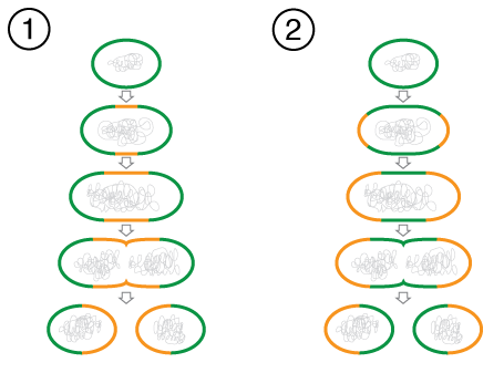
Most prokaryotic cells divide by the process of binary fission. A bacterial cell dividing this way is depicted in Figure below.
 Binary Fission in a Bacterial Cell. Cell division is relatively simple in prokaryotic cells. The two cells are dividing by binary fission. Green and orange lines indicate old and newly-generated bacterial cell walls, respectively. Eventually the parent cell will pinch apart to form two identical daughter cells. Left, growth at the center of bacterial body. Right, apical growth from the ends of the bacterial body.
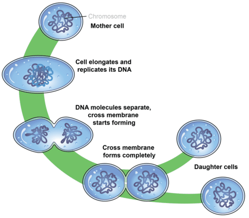
Binary fission can be described as a series of steps, although it is actually a continuous process. The steps are described below and also illustrated in Figure below. They include DNA replication, chromosome segregation, and finally the separation into two daughter cells.
- Step 1: DNA Replication. Just before the cell divides, its DNA is copied in a process called DNA replication. This results in two identical chromosomes instead of just one. This step is necessary so that when the cell divides, each daughter cell will have its own chromosome.
- Step 2: Chromosome Segregation. The two chromosomes segregate, or separate, and move to opposite ends (known as "poles") of the cell. This occurs as each copy of DNA attaches to different parts of the cell membrane.
- Step 3: Separation. A new plasma membrane starts growing into the center of the cell, and the cytoplasm splits apart, forming two daughter cells. As the cell begins to pull apart, the new and the original chromosomes are separated. The two daughter cells that result are genetically identical to each other and to the parent cell. New cell wall must also form around the two cells.

Steps of Binary Fission. Prokaryotic cells divide by binary fission. This is also how many single-celled organisms reproduce.
Cell Division in Eukaryotes
Cell division is more complex in eukaryotes than prokaryotes. Prior to dividing, all the DNA in a eukaryotic cell’s multiple chromosomes is replicated. Its organelles are also duplicated. Then, when the cell divides, it occurs in two major steps:
- The first step is mitosis, a multi-phase process in which the nucleus of the cell divides. During mitosis, the nuclear membrane breaks down and later reforms. The chromosomes are also sorted and separated to ensure that each daughter cell receives a diploid number (2 sets) of chromosomes. In humans, that number of chromosomes is 46 (23 pairs). Mitosis is described in greater detail in a subsequent concept.
- The second major step is cytokinesis. As in prokaryotic cells, the cytoplasm must divide. Cytokinesis is the division of the cytoplasm in eukaryotic cells, resulting in two genetically identical daughter cells.
Science Friday: The Axolotl: A Cut Above the Rest
The axolotl is an animal with the incredible ability to regenerate its legs. In this video by Science Friday, Dr. Susan Bryant discusses the research on how these animals are able to accomplish this amazing feat.
Summary
- Cell division is part of the life cycle of virtually all cells. Cell division is the process in which one cell divides to form two new cells.
- Most prokaryotic cells divide by the process of binary fission.
- In eukaryotes, cell division occurs in two major steps: mitosis and cytokinesis.
Review
- Describe binary fission.
- What is mitosis?
- Contrast cell division in prokaryotes and eukaryotes. Why are the two types of cell division different?
| Image | Reference | Attributions |
 |
[Figure 1] | Credit: Mariana Ruiz Villarreal (LadyofHats) for CK-12 Foundation;Ed Uthman Source: CK-12 Foundation ; CK12 ; web2.airmail.net/uthman/specimens/images/tvp.html License: CC BY-NC 3.0; Public Domain |
 |
[Figure 2] | Credit: Zachary Wilson;Mariana Ruiz Villarreal (LadyofHats) for CK-12 Foundation Source: CK-12 Foundation License: CC BY-NC 3.0 |
 |
[Figure 3] | Credit: Mariana Ruiz Villarreal (LadyofHats) for CK-12 Foundation Source: CK-12 Foundation License: CC BY-NC 3.0 |

