
1.6: Microscope
- Page ID
- 2935
\( \newcommand{\vecs}[1]{\overset { \scriptstyle \rightharpoonup} {\mathbf{#1}} } \)
\( \newcommand{\vecd}[1]{\overset{-\!-\!\rightharpoonup}{\vphantom{a}\smash {#1}}} \)
\( \newcommand{\id}{\mathrm{id}}\) \( \newcommand{\Span}{\mathrm{span}}\)
( \newcommand{\kernel}{\mathrm{null}\,}\) \( \newcommand{\range}{\mathrm{range}\,}\)
\( \newcommand{\RealPart}{\mathrm{Re}}\) \( \newcommand{\ImaginaryPart}{\mathrm{Im}}\)
\( \newcommand{\Argument}{\mathrm{Arg}}\) \( \newcommand{\norm}[1]{\| #1 \|}\)
\( \newcommand{\inner}[2]{\langle #1, #2 \rangle}\)
\( \newcommand{\Span}{\mathrm{span}}\)
\( \newcommand{\id}{\mathrm{id}}\)
\( \newcommand{\Span}{\mathrm{span}}\)
\( \newcommand{\kernel}{\mathrm{null}\,}\)
\( \newcommand{\range}{\mathrm{range}\,}\)
\( \newcommand{\RealPart}{\mathrm{Re}}\)
\( \newcommand{\ImaginaryPart}{\mathrm{Im}}\)
\( \newcommand{\Argument}{\mathrm{Arg}}\)
\( \newcommand{\norm}[1]{\| #1 \|}\)
\( \newcommand{\inner}[2]{\langle #1, #2 \rangle}\)
\( \newcommand{\Span}{\mathrm{span}}\) \( \newcommand{\AA}{\unicode[.8,0]{x212B}}\)
\( \newcommand{\vectorA}[1]{\vec{#1}} % arrow\)
\( \newcommand{\vectorAt}[1]{\vec{\text{#1}}} % arrow\)
\( \newcommand{\vectorB}[1]{\overset { \scriptstyle \rightharpoonup} {\mathbf{#1}} } \)
\( \newcommand{\vectorC}[1]{\textbf{#1}} \)
\( \newcommand{\vectorD}[1]{\overrightarrow{#1}} \)
\( \newcommand{\vectorDt}[1]{\overrightarrow{\text{#1}}} \)
\( \newcommand{\vectE}[1]{\overset{-\!-\!\rightharpoonup}{\vphantom{a}\smash{\mathbf {#1}}}} \)
\( \newcommand{\vecs}[1]{\overset { \scriptstyle \rightharpoonup} {\mathbf{#1}} } \)
\( \newcommand{\vecd}[1]{\overset{-\!-\!\rightharpoonup}{\vphantom{a}\smash {#1}}} \)
\(\newcommand{\avec}{\mathbf a}\) \(\newcommand{\bvec}{\mathbf b}\) \(\newcommand{\cvec}{\mathbf c}\) \(\newcommand{\dvec}{\mathbf d}\) \(\newcommand{\dtil}{\widetilde{\mathbf d}}\) \(\newcommand{\evec}{\mathbf e}\) \(\newcommand{\fvec}{\mathbf f}\) \(\newcommand{\nvec}{\mathbf n}\) \(\newcommand{\pvec}{\mathbf p}\) \(\newcommand{\qvec}{\mathbf q}\) \(\newcommand{\svec}{\mathbf s}\) \(\newcommand{\tvec}{\mathbf t}\) \(\newcommand{\uvec}{\mathbf u}\) \(\newcommand{\vvec}{\mathbf v}\) \(\newcommand{\wvec}{\mathbf w}\) \(\newcommand{\xvec}{\mathbf x}\) \(\newcommand{\yvec}{\mathbf y}\) \(\newcommand{\zvec}{\mathbf z}\) \(\newcommand{\rvec}{\mathbf r}\) \(\newcommand{\mvec}{\mathbf m}\) \(\newcommand{\zerovec}{\mathbf 0}\) \(\newcommand{\onevec}{\mathbf 1}\) \(\newcommand{\real}{\mathbb R}\) \(\newcommand{\twovec}[2]{\left[\begin{array}{r}#1 \\ #2 \end{array}\right]}\) \(\newcommand{\ctwovec}[2]{\left[\begin{array}{c}#1 \\ #2 \end{array}\right]}\) \(\newcommand{\threevec}[3]{\left[\begin{array}{r}#1 \\ #2 \\ #3 \end{array}\right]}\) \(\newcommand{\cthreevec}[3]{\left[\begin{array}{c}#1 \\ #2 \\ #3 \end{array}\right]}\) \(\newcommand{\fourvec}[4]{\left[\begin{array}{r}#1 \\ #2 \\ #3 \\ #4 \end{array}\right]}\) \(\newcommand{\cfourvec}[4]{\left[\begin{array}{c}#1 \\ #2 \\ #3 \\ #4 \end{array}\right]}\) \(\newcommand{\fivevec}[5]{\left[\begin{array}{r}#1 \\ #2 \\ #3 \\ #4 \\ #5 \\ \end{array}\right]}\) \(\newcommand{\cfivevec}[5]{\left[\begin{array}{c}#1 \\ #2 \\ #3 \\ #4 \\ #5 \\ \end{array}\right]}\) \(\newcommand{\mattwo}[4]{\left[\begin{array}{rr}#1 \amp #2 \\ #3 \amp #4 \\ \end{array}\right]}\) \(\newcommand{\laspan}[1]{\text{Span}\{#1\}}\) \(\newcommand{\bcal}{\cal B}\) \(\newcommand{\ccal}{\cal C}\) \(\newcommand{\scal}{\cal S}\) \(\newcommand{\wcal}{\cal W}\) \(\newcommand{\ecal}{\cal E}\) \(\newcommand{\coords}[2]{\left\{#1\right\}_{#2}}\) \(\newcommand{\gray}[1]{\color{gray}{#1}}\) \(\newcommand{\lgray}[1]{\color{lightgray}{#1}}\) \(\newcommand{\rank}{\operatorname{rank}}\) \(\newcommand{\row}{\text{Row}}\) \(\newcommand{\col}{\text{Col}}\) \(\renewcommand{\row}{\text{Row}}\) \(\newcommand{\nul}{\text{Nul}}\) \(\newcommand{\var}{\text{Var}}\) \(\newcommand{\corr}{\text{corr}}\) \(\newcommand{\len}[1]{\left|#1\right|}\) \(\newcommand{\bbar}{\overline{\bvec}}\) \(\newcommand{\bhat}{\widehat{\bvec}}\) \(\newcommand{\bperp}{\bvec^\perp}\) \(\newcommand{\xhat}{\widehat{\xvec}}\) \(\newcommand{\vhat}{\widehat{\vvec}}\) \(\newcommand{\uhat}{\widehat{\uvec}}\) \(\newcommand{\what}{\widehat{\wvec}}\) \(\newcommand{\Sighat}{\widehat{\Sigma}}\) \(\newcommand{\lt}{<}\) \(\newcommand{\gt}{>}\) \(\newcommand{\amp}{&}\) \(\definecolor{fillinmathshade}{gray}{0.9}\)
How can we see the details of bacteria?
With the naked eye, bacteria just look like a slimy smear on a petri dish. How can we study them in more detail? The invention of the microscope has allowed us to see bacteria, cells, and other things too small to be seen with the naked eye.
The Microscope
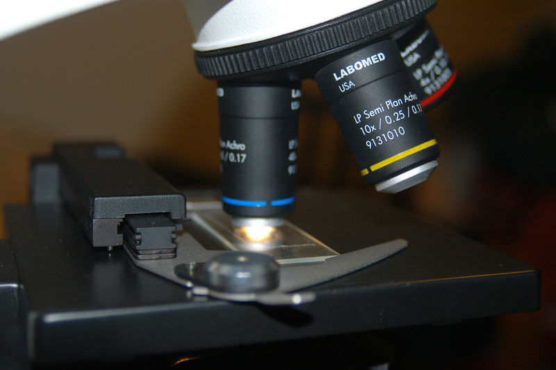
Microscopes, tools that you may get to use in your class, are some of the most important tools in biology (Figure below). A microscope is a tool used to make things that are too small to be seen by the human eye look bigger. Microscopy is the study of small objects using microscopes. Look at your fingertips. Before microscopes were invented in 1595, the smallest things you could see on yourself were the tiny lines in your skin. But what else is hidden in your skin?

Invention of the Microscope
Over four hundred years ago, two Dutch spectacle makers, Zacharias Janssen and his son Hans, were experimenting with several lenses in a tube. They discovered that nearby objects appeared greatly enlarged, or magnified. This was the forerunner of the compound microscope and of the telescope.
In 1665, Robert Hooke, an English natural scientist, used a microscope to zoom in on a piece of cork - the stuff that makes up the stoppers in wine bottles, which is made from tree bark. Inside of cork, he discovered tiny structures, which he called cells. It turns out that cells are the smallest structural unit of living organisms. This finding eventually led to the development of the theory that all living things are made of cells. Without microscopes, this discovery would not have been possible, and the cell theory would not have been developed.
Hooke's discovery of the cell set the stage for other scientists to discover other types of organisms. After Hooke, the "father of microscopy," Dutch scientist Antonie van Leeuwenhoek (Figure below) taught himself to make one of the first microscopes. In one of his early experiments, van Leeuwenhoek took a sample of scum from his own teeth and used his microscope to discover bacteria, the smallest living organism on the planet. Using microscopes, van Leeuwenhoek also discovered one-celled protists and sperm cells.
Today, microscopes are used by all types of scientists, including cell biologists, microbiologists, virologists, forensic scientists, entomologists, taxonomists, and many other types.

Types of Microscopes
Some modern microscopes use light, as Hooke's and van Leeuwenhoek's did. Others may use electron beams or sound waves. Researchers now use these four types of microscopes:
- Light microscopes allow biologists to see small details of a specimen. Most of the microscopes used in schools and laboratories are light microscopes. Light microscopes use lenses, typically made of glass or plastic, to focus light either into the eye, a camera, or some other light detector. The most powerful light microscopes can make images up to 2,000 times larger.
- Transmission electron microscopes (TEM) focus a beam of electrons through an object and can make an image up to two million times bigger, with a very clear image.
- Scanning electron microscopes (SEM) allow scientists to find the shape and surface texture of extremely small objects, including a paperclip, a bedbug, or even an atom. These microscopes slide a beam of electrons across the surface of a specimen, producing detailed maps of the surface of objects. Magnification in a SEM can be controlled over a range from about 10 to 500,000 times.
- Scanning acoustic microscopes use sound waves to scan a specimen. These microscopes are useful in biology and medical research.

Science Friday: Stained Glass Conservation
Stained glass from the Middle Ages is often hundreds of years old. Unfortunately, many of these relics are in need of cleaning and maintenance. In this video by Science Friday, conservator Mary Higgins discusses the methods used to protect the stained glass.
Summary
- A microscope is a tool used to make things that are too small to be seen by the naked eye look bigger.
- Types of microscopes include light microscopes, transmission electron microscopes (TEM), scanning electron microscopes (SEM), and scanning acoustic microscopes.
Explore More
Use the resources below to answer the questions that follow.
- Using a microscope at http://www.youtube.com/watch?v=bGBgABLEV4g (4:01)
- How should you carry a compound optical microscope?
- What procedure should you use when seeking to use the most powerful optical lenses?
Review
- What is the purpose of a microscope?
- What were the findings of Hooke and van Leeuwenhoek?
- What are the differences between a light microscope and a scanning electron microscope?