2.11: Parts of the Cell
- Page ID
- 1439
\( \newcommand{\vecs}[1]{\overset { \scriptstyle \rightharpoonup} {\mathbf{#1}} } \)
\( \newcommand{\vecd}[1]{\overset{-\!-\!\rightharpoonup}{\vphantom{a}\smash {#1}}} \)
\( \newcommand{\dsum}{\displaystyle\sum\limits} \)
\( \newcommand{\dint}{\displaystyle\int\limits} \)
\( \newcommand{\dlim}{\displaystyle\lim\limits} \)
\( \newcommand{\id}{\mathrm{id}}\) \( \newcommand{\Span}{\mathrm{span}}\)
( \newcommand{\kernel}{\mathrm{null}\,}\) \( \newcommand{\range}{\mathrm{range}\,}\)
\( \newcommand{\RealPart}{\mathrm{Re}}\) \( \newcommand{\ImaginaryPart}{\mathrm{Im}}\)
\( \newcommand{\Argument}{\mathrm{Arg}}\) \( \newcommand{\norm}[1]{\| #1 \|}\)
\( \newcommand{\inner}[2]{\langle #1, #2 \rangle}\)
\( \newcommand{\Span}{\mathrm{span}}\)
\( \newcommand{\id}{\mathrm{id}}\)
\( \newcommand{\Span}{\mathrm{span}}\)
\( \newcommand{\kernel}{\mathrm{null}\,}\)
\( \newcommand{\range}{\mathrm{range}\,}\)
\( \newcommand{\RealPart}{\mathrm{Re}}\)
\( \newcommand{\ImaginaryPart}{\mathrm{Im}}\)
\( \newcommand{\Argument}{\mathrm{Arg}}\)
\( \newcommand{\norm}[1]{\| #1 \|}\)
\( \newcommand{\inner}[2]{\langle #1, #2 \rangle}\)
\( \newcommand{\Span}{\mathrm{span}}\) \( \newcommand{\AA}{\unicode[.8,0]{x212B}}\)
\( \newcommand{\vectorA}[1]{\vec{#1}} % arrow\)
\( \newcommand{\vectorAt}[1]{\vec{\text{#1}}} % arrow\)
\( \newcommand{\vectorB}[1]{\overset { \scriptstyle \rightharpoonup} {\mathbf{#1}} } \)
\( \newcommand{\vectorC}[1]{\textbf{#1}} \)
\( \newcommand{\vectorD}[1]{\overrightarrow{#1}} \)
\( \newcommand{\vectorDt}[1]{\overrightarrow{\text{#1}}} \)
\( \newcommand{\vectE}[1]{\overset{-\!-\!\rightharpoonup}{\vphantom{a}\smash{\mathbf {#1}}}} \)
\( \newcommand{\vecs}[1]{\overset { \scriptstyle \rightharpoonup} {\mathbf{#1}} } \)
\(\newcommand{\longvect}{\overrightarrow}\)
\( \newcommand{\vecd}[1]{\overset{-\!-\!\rightharpoonup}{\vphantom{a}\smash {#1}}} \)
\(\newcommand{\avec}{\mathbf a}\) \(\newcommand{\bvec}{\mathbf b}\) \(\newcommand{\cvec}{\mathbf c}\) \(\newcommand{\dvec}{\mathbf d}\) \(\newcommand{\dtil}{\widetilde{\mathbf d}}\) \(\newcommand{\evec}{\mathbf e}\) \(\newcommand{\fvec}{\mathbf f}\) \(\newcommand{\nvec}{\mathbf n}\) \(\newcommand{\pvec}{\mathbf p}\) \(\newcommand{\qvec}{\mathbf q}\) \(\newcommand{\svec}{\mathbf s}\) \(\newcommand{\tvec}{\mathbf t}\) \(\newcommand{\uvec}{\mathbf u}\) \(\newcommand{\vvec}{\mathbf v}\) \(\newcommand{\wvec}{\mathbf w}\) \(\newcommand{\xvec}{\mathbf x}\) \(\newcommand{\yvec}{\mathbf y}\) \(\newcommand{\zvec}{\mathbf z}\) \(\newcommand{\rvec}{\mathbf r}\) \(\newcommand{\mvec}{\mathbf m}\) \(\newcommand{\zerovec}{\mathbf 0}\) \(\newcommand{\onevec}{\mathbf 1}\) \(\newcommand{\real}{\mathbb R}\) \(\newcommand{\twovec}[2]{\left[\begin{array}{r}#1 \\ #2 \end{array}\right]}\) \(\newcommand{\ctwovec}[2]{\left[\begin{array}{c}#1 \\ #2 \end{array}\right]}\) \(\newcommand{\threevec}[3]{\left[\begin{array}{r}#1 \\ #2 \\ #3 \end{array}\right]}\) \(\newcommand{\cthreevec}[3]{\left[\begin{array}{c}#1 \\ #2 \\ #3 \end{array}\right]}\) \(\newcommand{\fourvec}[4]{\left[\begin{array}{r}#1 \\ #2 \\ #3 \\ #4 \end{array}\right]}\) \(\newcommand{\cfourvec}[4]{\left[\begin{array}{c}#1 \\ #2 \\ #3 \\ #4 \end{array}\right]}\) \(\newcommand{\fivevec}[5]{\left[\begin{array}{r}#1 \\ #2 \\ #3 \\ #4 \\ #5 \\ \end{array}\right]}\) \(\newcommand{\cfivevec}[5]{\left[\begin{array}{c}#1 \\ #2 \\ #3 \\ #4 \\ #5 \\ \end{array}\right]}\) \(\newcommand{\mattwo}[4]{\left[\begin{array}{rr}#1 \amp #2 \\ #3 \amp #4 \\ \end{array}\right]}\) \(\newcommand{\laspan}[1]{\text{Span}\{#1\}}\) \(\newcommand{\bcal}{\cal B}\) \(\newcommand{\ccal}{\cal C}\) \(\newcommand{\scal}{\cal S}\) \(\newcommand{\wcal}{\cal W}\) \(\newcommand{\ecal}{\cal E}\) \(\newcommand{\coords}[2]{\left\{#1\right\}_{#2}}\) \(\newcommand{\gray}[1]{\color{gray}{#1}}\) \(\newcommand{\lgray}[1]{\color{lightgray}{#1}}\) \(\newcommand{\rank}{\operatorname{rank}}\) \(\newcommand{\row}{\text{Row}}\) \(\newcommand{\col}{\text{Col}}\) \(\renewcommand{\row}{\text{Row}}\) \(\newcommand{\nul}{\text{Nul}}\) \(\newcommand{\var}{\text{Var}}\) \(\newcommand{\corr}{\text{corr}}\) \(\newcommand{\len}[1]{\left|#1\right|}\) \(\newcommand{\bbar}{\overline{\bvec}}\) \(\newcommand{\bhat}{\widehat{\bvec}}\) \(\newcommand{\bperp}{\bvec^\perp}\) \(\newcommand{\xhat}{\widehat{\xvec}}\) \(\newcommand{\vhat}{\widehat{\vvec}}\) \(\newcommand{\uhat}{\widehat{\uvec}}\) \(\newcommand{\what}{\widehat{\wvec}}\) \(\newcommand{\Sighat}{\widehat{\Sigma}}\) \(\newcommand{\lt}{<}\) \(\newcommand{\gt}{>}\) \(\newcommand{\amp}{&}\) \(\definecolor{fillinmathshade}{gray}{0.9}\)
What do a bacterial cell and one of your cells have in common?
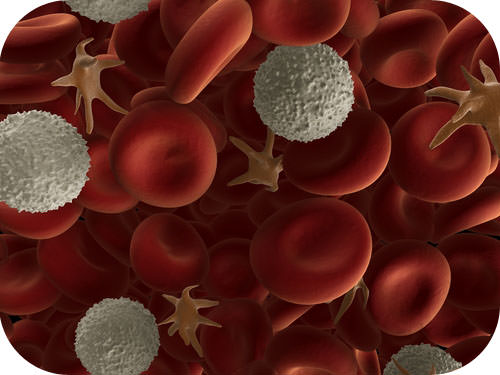
There are many different types of cells, but they all have certain parts in common. As this image of human blood shows, cells come in different shapes and sizes. The shapes and sizes directly influence the function of the cell. Yet, all cells - cells from the smallest bacteria to those in the largest whale - do some similar functions, so they do have parts in common.
Common Parts of Cells
Discovery of Cells
The first time the word cell was used to refer to these tiny units of life was in 1665 by a British scientist named Robert Hooke. Hooke was one of the earliest scientists to study living things under a microscope. The microscopes of his day were not very strong, but Hooke was still able to make an important discovery. When he looked at a thin slice of cork under his microscope, he was surprised to see what looked like a honeycomb. Hooke made the drawing in the Figure below to show what he saw. As you can see, the cork was made up of many tiny units, which Hooke called cells.
 Cork Cells. This is what Robert Hooke saw when he looked at a thin slice of cork under his microscope. What type of material is cork? Do you know where cork comes from?
Soon after Robert Hooke discovered cells in cork, Anton van Leeuwenhoek in Holland made other important discoveries using a microscope. Leeuwenhoek made his own microscope lenses, and he was so good at it that his microscope was more powerful than other microscopes of his day. In fact, Leeuwenhoek’s microscope was almost as strong as modern light microscopes.
Using his microscope, Leeuwenhoek discovered tiny animals such as rotifers. Leeuwenhoek also discovered human blood cells. He even scraped plaque from his own teeth and observed it under the microscope. What do you think Leeuwenhoek saw in the plaque? He saw tiny living things with a single cell that he named animalcules (“tiny animals”). Today, we call Leeuwenhoek’s animalcules bacteria.
The Cell Theory
The Cell Theory is one of the fundamental theories of biology. For two centuries after the discovery of the microscope by Robert Hooke and Anton van Leeuwenhoek, biologists found cells everywhere. Biologists in the early part of the 19th century suggested that all living things were made of cells, but the role of cells as the primary building block of life was not discovered until 1839 when two German scientists, Theodor Schwann, a zoologist (studies animals), and Matthias Jakob Schleiden, a botanist (studies plants), suggested that cells were the basic unit of structure and function of all life. Later, in 1858, the German doctor Rudolf Virchow observed that cells divide to produce more cells. He proposed that all cells arise only from other cells. The collective observations of all three scientists form the Cell Theory, which states that:
- all organisms are made up of one or more cells,
- all the life functions of an organism occur within cells,
- all cells come from preexisting cells.
Cell Diversity
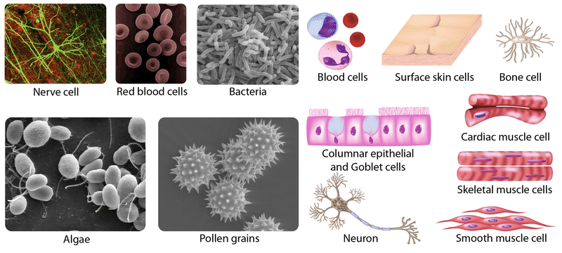
Cells with different functions often have different shapes. The cells pictured in the Figure below are just a few examples of the many different shapes that cells may have. Each type of cell in the figure has a shape that helps it do its job. For example, the job of the nerve cell is to carry messages to other cells. The nerve cell has many long extensions that reach out in all directions, allowing it to pass messages to many other cells at once. Do you see the tail-like projections on the algae cells? Algae live in water, and their tails help them swim. Pollen grains have spikes that help them stick to insects such as bees. How do you think the spikes help the pollen grains do their job? (Hint: Insects pollinate flowers.)
 As these pictures show, cells come in many different shapes. How are the shapes of these cells related to their functions?
Four Common Parts of a Cell
Although cells are diverse, all cells have certain parts in common. The parts include a plasma membrane, cytoplasm, ribosomes, and DNA.
- The plasma membrane (also called the cell membrane) is a thin coat of lipids that surrounds a cell. It forms the physical boundary between the cell and its environment, so you can think of it as the ‘‘skin’’ of the cell.
- Cytoplasm refers to all of the cellular material inside the plasma membrane, other than the nucleus. Cytoplasm is made up of a watery substance called cytosol, and contains other cell structures such as ribosomes.
- Ribosomes are structures in the cytoplasm where proteins are made.
- DNA is a nucleic acid found in cells. It contains the genetic instructions that cells need to make proteins.
These parts are common to all cells, from organisms as different as bacteria and human beings. How did all known organisms come to have such similar cells? The similarities show that all life on Earth has a common evolutionary history.
Summary
- Cells come in many different shapes. Cells with different functions often have different shapes.
- Although cells comes in diverse shapes, all cells have certain parts in common. These parts include the plasma membrane, cytoplasm, ribosomes, and DNA.
Review
- Who coined the term cell, in reference to the tiny structures seen in living organisms?
- Who identified animalcules? What are animalcules?
- What are the three main parts of the cell theory?
- List the four parts common to all cells.
- What are the cell structures where proteins are made?
- What is the role of DNA?
| Image | Reference | Attributions |
 |
[Figure 1] | Credit: Nerve cell: WA Lee et al.; Blood cell: Courtesy of National Institute of Health; Bacteria: TJ Kirn, MJ Lafferty, CMP Sandoe, and RK Taylor; Algae: EF Smith and PA Lefebvre; Pollen: L Howard and C Daghlian; Illustrations: Image copyright Alila Medical Media, 2014 Source: Nerve cell: commons.wikimedia.org/wiki/File:GFPneuron.png ; Blood cell: commons.wikimedia.org/wiki/File:Redbloodcells.jpg ; Bacteria: remf.dartmouth.edu/images/bacteriaSEM/source/1.html ; Algae: remf.dartmouth.edu/images/algaeSEM/source/1.html ; Pollen: remf.dartmouth.edu/images/botanicalPollenSEM/source/10.html ; Illustrations: http://www.shutterstock.com License: (Nerve cel) CC BY 2.5; (Blood cell) Public Domain; (Bacteria) Public Domain; (Algae) Public Domain; (Pollen) Public Domain; (Illustrations) License from Shutterstock |
 |
[Figure 2] | Credit: Cork cell: Robert Hooke, Micrographia,1665; Tree branch: OpenClips Source: Cork cell: commons.wikimedia.org/wiki/Image:RobertHookeMicrographia1665.jpg ; Tree branch: pixabay.com/en/birch-branch-leaves-plant-nature-155882/ License: Public Domain |
 |
[Figure 3] | Credit: Nerve cell: WA Lee et al.; Blood cell: Courtesy of National Institute of Health; Bacteria: TJ Kirn, MJ Lafferty, CMP Sandoe, and RK Taylor; Algae: EF Smith and PA Lefebvre; Pollen: L Howard and C Daghlian; Illustrations: Image copyright Alila Medical Media, 2014 Source: Nerve cell: commons.wikimedia.org/wiki/File:GFPneuron.png ; Blood cell: commons.wikimedia.org/wiki/File:Redbloodcells.jpg ; Bacteria: remf.dartmouth.edu/images/bacteriaSEM/source/1.html ; Algae: remf.dartmouth.edu/images/algaeSEM/source/1.html ; Pollen: remf.dartmouth.edu/images/botanicalPollenSEM/source/10.html ; Illustrations: http://www.shutterstock.com License: (Nerve cel) CC BY 2.5; (Blood cell) Public Domain; (Bacteria) Public Domain; (Algae) Public Domain; (Pollen) Public Domain; (Illustrations) License from Shutterstock |

