
7.1: Virus Structure
- Page ID
- 12185
\( \newcommand{\vecs}[1]{\overset { \scriptstyle \rightharpoonup} {\mathbf{#1}} } \)
\( \newcommand{\vecd}[1]{\overset{-\!-\!\rightharpoonup}{\vphantom{a}\smash {#1}}} \)
\( \newcommand{\dsum}{\displaystyle\sum\limits} \)
\( \newcommand{\dint}{\displaystyle\int\limits} \)
\( \newcommand{\dlim}{\displaystyle\lim\limits} \)
\( \newcommand{\id}{\mathrm{id}}\) \( \newcommand{\Span}{\mathrm{span}}\)
( \newcommand{\kernel}{\mathrm{null}\,}\) \( \newcommand{\range}{\mathrm{range}\,}\)
\( \newcommand{\RealPart}{\mathrm{Re}}\) \( \newcommand{\ImaginaryPart}{\mathrm{Im}}\)
\( \newcommand{\Argument}{\mathrm{Arg}}\) \( \newcommand{\norm}[1]{\| #1 \|}\)
\( \newcommand{\inner}[2]{\langle #1, #2 \rangle}\)
\( \newcommand{\Span}{\mathrm{span}}\)
\( \newcommand{\id}{\mathrm{id}}\)
\( \newcommand{\Span}{\mathrm{span}}\)
\( \newcommand{\kernel}{\mathrm{null}\,}\)
\( \newcommand{\range}{\mathrm{range}\,}\)
\( \newcommand{\RealPart}{\mathrm{Re}}\)
\( \newcommand{\ImaginaryPart}{\mathrm{Im}}\)
\( \newcommand{\Argument}{\mathrm{Arg}}\)
\( \newcommand{\norm}[1]{\| #1 \|}\)
\( \newcommand{\inner}[2]{\langle #1, #2 \rangle}\)
\( \newcommand{\Span}{\mathrm{span}}\) \( \newcommand{\AA}{\unicode[.8,0]{x212B}}\)
\( \newcommand{\vectorA}[1]{\vec{#1}} % arrow\)
\( \newcommand{\vectorAt}[1]{\vec{\text{#1}}} % arrow\)
\( \newcommand{\vectorB}[1]{\overset { \scriptstyle \rightharpoonup} {\mathbf{#1}} } \)
\( \newcommand{\vectorC}[1]{\textbf{#1}} \)
\( \newcommand{\vectorD}[1]{\overrightarrow{#1}} \)
\( \newcommand{\vectorDt}[1]{\overrightarrow{\text{#1}}} \)
\( \newcommand{\vectE}[1]{\overset{-\!-\!\rightharpoonup}{\vphantom{a}\smash{\mathbf {#1}}}} \)
\( \newcommand{\vecs}[1]{\overset { \scriptstyle \rightharpoonup} {\mathbf{#1}} } \)
\(\newcommand{\longvect}{\overrightarrow}\)
\( \newcommand{\vecd}[1]{\overset{-\!-\!\rightharpoonup}{\vphantom{a}\smash {#1}}} \)
\(\newcommand{\avec}{\mathbf a}\) \(\newcommand{\bvec}{\mathbf b}\) \(\newcommand{\cvec}{\mathbf c}\) \(\newcommand{\dvec}{\mathbf d}\) \(\newcommand{\dtil}{\widetilde{\mathbf d}}\) \(\newcommand{\evec}{\mathbf e}\) \(\newcommand{\fvec}{\mathbf f}\) \(\newcommand{\nvec}{\mathbf n}\) \(\newcommand{\pvec}{\mathbf p}\) \(\newcommand{\qvec}{\mathbf q}\) \(\newcommand{\svec}{\mathbf s}\) \(\newcommand{\tvec}{\mathbf t}\) \(\newcommand{\uvec}{\mathbf u}\) \(\newcommand{\vvec}{\mathbf v}\) \(\newcommand{\wvec}{\mathbf w}\) \(\newcommand{\xvec}{\mathbf x}\) \(\newcommand{\yvec}{\mathbf y}\) \(\newcommand{\zvec}{\mathbf z}\) \(\newcommand{\rvec}{\mathbf r}\) \(\newcommand{\mvec}{\mathbf m}\) \(\newcommand{\zerovec}{\mathbf 0}\) \(\newcommand{\onevec}{\mathbf 1}\) \(\newcommand{\real}{\mathbb R}\) \(\newcommand{\twovec}[2]{\left[\begin{array}{r}#1 \\ #2 \end{array}\right]}\) \(\newcommand{\ctwovec}[2]{\left[\begin{array}{c}#1 \\ #2 \end{array}\right]}\) \(\newcommand{\threevec}[3]{\left[\begin{array}{r}#1 \\ #2 \\ #3 \end{array}\right]}\) \(\newcommand{\cthreevec}[3]{\left[\begin{array}{c}#1 \\ #2 \\ #3 \end{array}\right]}\) \(\newcommand{\fourvec}[4]{\left[\begin{array}{r}#1 \\ #2 \\ #3 \\ #4 \end{array}\right]}\) \(\newcommand{\cfourvec}[4]{\left[\begin{array}{c}#1 \\ #2 \\ #3 \\ #4 \end{array}\right]}\) \(\newcommand{\fivevec}[5]{\left[\begin{array}{r}#1 \\ #2 \\ #3 \\ #4 \\ #5 \\ \end{array}\right]}\) \(\newcommand{\cfivevec}[5]{\left[\begin{array}{c}#1 \\ #2 \\ #3 \\ #4 \\ #5 \\ \end{array}\right]}\) \(\newcommand{\mattwo}[4]{\left[\begin{array}{rr}#1 \amp #2 \\ #3 \amp #4 \\ \end{array}\right]}\) \(\newcommand{\laspan}[1]{\text{Span}\{#1\}}\) \(\newcommand{\bcal}{\cal B}\) \(\newcommand{\ccal}{\cal C}\) \(\newcommand{\scal}{\cal S}\) \(\newcommand{\wcal}{\cal W}\) \(\newcommand{\ecal}{\cal E}\) \(\newcommand{\coords}[2]{\left\{#1\right\}_{#2}}\) \(\newcommand{\gray}[1]{\color{gray}{#1}}\) \(\newcommand{\lgray}[1]{\color{lightgray}{#1}}\) \(\newcommand{\rank}{\operatorname{rank}}\) \(\newcommand{\row}{\text{Row}}\) \(\newcommand{\col}{\text{Col}}\) \(\renewcommand{\row}{\text{Row}}\) \(\newcommand{\nul}{\text{Nul}}\) \(\newcommand{\var}{\text{Var}}\) \(\newcommand{\corr}{\text{corr}}\) \(\newcommand{\len}[1]{\left|#1\right|}\) \(\newcommand{\bbar}{\overline{\bvec}}\) \(\newcommand{\bhat}{\widehat{\bvec}}\) \(\newcommand{\bperp}{\bvec^\perp}\) \(\newcommand{\xhat}{\widehat{\xvec}}\) \(\newcommand{\vhat}{\widehat{\vvec}}\) \(\newcommand{\uhat}{\widehat{\uvec}}\) \(\newcommand{\what}{\widehat{\wvec}}\) \(\newcommand{\Sighat}{\widehat{\Sigma}}\) \(\newcommand{\lt}{<}\) \(\newcommand{\gt}{>}\) \(\newcommand{\amp}{&}\) \(\definecolor{fillinmathshade}{gray}{0.9}\)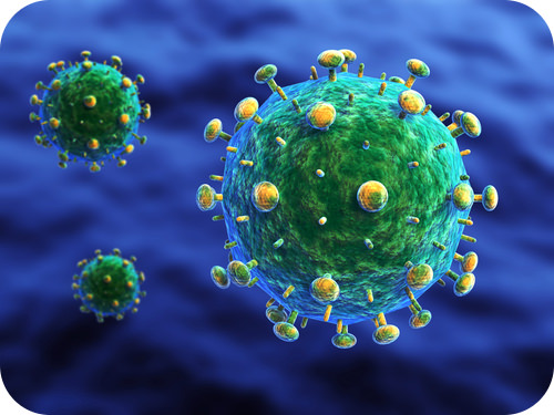
Is this a cell or a virus?
It is actually a representation of the HIV virus, the virus that causes AIDS. All the little “knobs” on the outside of the virus help to give the virus structure. And it is this structure that must be identified by a vaccine.
Structure of Viruses
Viruses vary in their structure. A virus particle consists of DNA or RNA within a protective protein coat called a capsid. The shape of the capsid may vary from one type of virus to another. The capsid is made from the proteins that are encoded by viral genes within their genome.
The shape of the capsid serves as one basis for classification of viruses. The capsid of the virus shown in Figure below is icosahedral. Virally coded proteins will self-assemble to form a capsid. Some viruses have an envelope of phospholipids and proteins. The envelope is made from portions of the host’s cell membrane. It surrounds the capsid and helps protect the virus from the host’s immune system. The envelope may also have receptor molecules that can bind with host cells. They make it easier for the virus to infect the cells.
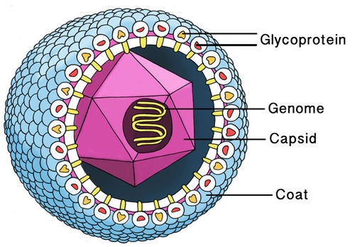 Diagram of a Cytomegalovirus. The capsid encloses the genetic material of the virus. The envelope which surrounds the capsid is typically made from portions of the host cell membranes (phospholipids and proteins). Not all viruses have a viral envelope.
Helical Viruses
Helical capsids are made up of a single type of protein subunit stacked around a central axis to form a helical structure. The helix may have a hollow center, which makes it look like a hollow tube. This arrangement results in rod-shaped or filamentous virions. These virions can be anything from short and very rigid, to long and very flexible. The well-studied tobacco mosaic virus (TMV) is an example of a helical virus, as seen in the Figure below.
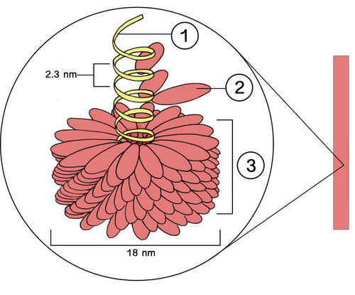
A helical virus, tobacco mosaic virus. Although their diameter may be very small, some helical viruses can be quite long, as shown here. 1. Nucleic acid; 2. Viral protein units, 3. Capsid. TMV causes tobacco mosaic disease in tobacco, cucumber, pepper, and tomato plants.
Icosahedral Viruses
Icosahedral capsid symmetry gives viruses a spherical appearance at low magnification, but the protein subunits are actually arranged in a regular geometrical pattern, similar to a soccer ball; they are not truly spherical. An icosahedral shape is the most efficient way of creating a hardy structure from multiple copies of a single protein. This shape is used because it can be built from a single basic unit protein which is used over and over again. This saves space in the viral genome.
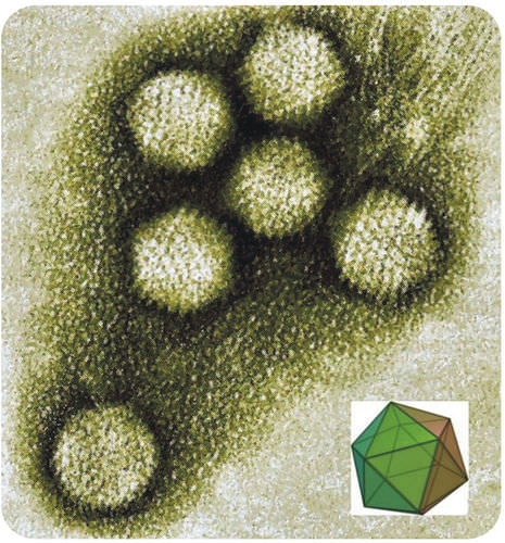 Adenovirus, an icosahedral virus. An icosahedron is a three-dimensional shape made up of 20 equilateral triangles. Viral structures are built of repeated identical protein subunits, making the icosahedron the easiest shape to assemble using these subunits.
Complex Viruses
Complex viruses possess a capsid which is neither purely helical, nor purely icosahedral, and which may have extra structures such as protein tails or a complex outer wall. Viral protein subunits will self-assemble into a capsid, but the complex viruses DNA also codes for proteins which help in building the viral capsid. Many phage viruses are complex-shaped; they have an icosahedral head bound to a helical tail. The tail may have a base plate with protein tail fibers. Some complex viruses do not have tail fibers.
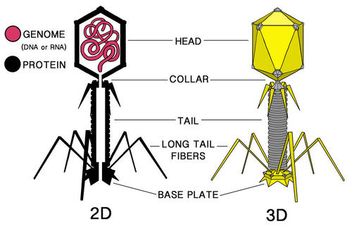 This “moon lander”-shaped complex virus infects Escherichia coli bacteria.
Enveloped Viruses
Some viruses are able to surround (envelop) themselves in a portion of the cell membrane of their host. The virus can use either the outer membrane of the host cell, or an internal membrane such as the nuclear membrane or endoplasmic reticulum. In this way the virus gains an outer lipid bilayer known as a viral envelope. This membrane is studded with proteins coded for by both the viral genome and the host genome. However, the lipid membrane itself and any carbohydrates present come entirely from the host cell. The influenza virus, HIV, and the varicella zoster virus (Figure below) are enveloped viruses.
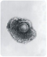 An enveloped virus. Varicella zoster virus causes chicken pox and shingles.
The viral envelope can give a virus some advantages over other capsid-only viruses. For example, they have better protection from the host's immune system, enzymes and certain chemicals. The proteins in the envelope can include glycoproteins, which act as receptor molecules. These receptor molecules allow host cells to recognize and bind the virions, which may result in easier uptake of the virion into the cell. Most enveloped viruses depend on their envelopes to infect cells. However, because the envelope contains lipids, it makes the virus more susceptible to inactivation by environmental agents, such as detergents that disrupt lipids.
Summary
- Viruses have different shapes. They can be cylindrical, icosahedral, complex, or enveloped.
Review
- Describe variation in capsid shape in viruses.
- Compare the structures of a prokaryote and a virus. If you prefer, you may draw a diagram of each and label the different parts of each structure.
| Image | Reference | Attributions |
|
[Figure 1] | Credit: Courtesy of Centers for Disease Control and Prevention/Dr. Erskine Palmer/B.G. Partin Source: commons.wikimedia.org/wiki/File:Varicella_%28Chickenpox%29_Virus_PHIL_1878_lores.jpg License: Public Domain |
 |
[Figure 2] | Credit: Laura Guerin Source: CK-12 Foundation License: CC BY-NC 3.0 |
 |
[Figure 3] | Credit: Laura Guerin Source: CK-12 Foundation License: CC BY-NC 3.0 |
 |
[Figure 4] | Credit: User:GrahamColm/Wikipedia Source: en.Wikipedia.org/wiki/Image:Enteric_Adenoviruses.jpg License: Public Domain |
 |
[Figure 5] | Credit: Laura Guerin Source: CK-12 Foundation License: CC BY-NC 3.0 |
 |
[Figure 6] | Credit: Courtesy of Centers for Disease Control and Prevention/Dr. Erskine Palmer/B.G. Partin Source: commons.wikimedia.org/wiki/File:Varicella_%28Chickenpox%29_Virus_PHIL_1878_lores.jpg License: Public Domain |