13.11: Human Body
- Page ID
- 1395
\( \newcommand{\vecs}[1]{\overset { \scriptstyle \rightharpoonup} {\mathbf{#1}} } \)
\( \newcommand{\vecd}[1]{\overset{-\!-\!\rightharpoonup}{\vphantom{a}\smash {#1}}} \)
\( \newcommand{\dsum}{\displaystyle\sum\limits} \)
\( \newcommand{\dint}{\displaystyle\int\limits} \)
\( \newcommand{\dlim}{\displaystyle\lim\limits} \)
\( \newcommand{\id}{\mathrm{id}}\) \( \newcommand{\Span}{\mathrm{span}}\)
( \newcommand{\kernel}{\mathrm{null}\,}\) \( \newcommand{\range}{\mathrm{range}\,}\)
\( \newcommand{\RealPart}{\mathrm{Re}}\) \( \newcommand{\ImaginaryPart}{\mathrm{Im}}\)
\( \newcommand{\Argument}{\mathrm{Arg}}\) \( \newcommand{\norm}[1]{\| #1 \|}\)
\( \newcommand{\inner}[2]{\langle #1, #2 \rangle}\)
\( \newcommand{\Span}{\mathrm{span}}\)
\( \newcommand{\id}{\mathrm{id}}\)
\( \newcommand{\Span}{\mathrm{span}}\)
\( \newcommand{\kernel}{\mathrm{null}\,}\)
\( \newcommand{\range}{\mathrm{range}\,}\)
\( \newcommand{\RealPart}{\mathrm{Re}}\)
\( \newcommand{\ImaginaryPart}{\mathrm{Im}}\)
\( \newcommand{\Argument}{\mathrm{Arg}}\)
\( \newcommand{\norm}[1]{\| #1 \|}\)
\( \newcommand{\inner}[2]{\langle #1, #2 \rangle}\)
\( \newcommand{\Span}{\mathrm{span}}\) \( \newcommand{\AA}{\unicode[.8,0]{x212B}}\)
\( \newcommand{\vectorA}[1]{\vec{#1}} % arrow\)
\( \newcommand{\vectorAt}[1]{\vec{\text{#1}}} % arrow\)
\( \newcommand{\vectorB}[1]{\overset { \scriptstyle \rightharpoonup} {\mathbf{#1}} } \)
\( \newcommand{\vectorC}[1]{\textbf{#1}} \)
\( \newcommand{\vectorD}[1]{\overrightarrow{#1}} \)
\( \newcommand{\vectorDt}[1]{\overrightarrow{\text{#1}}} \)
\( \newcommand{\vectE}[1]{\overset{-\!-\!\rightharpoonup}{\vphantom{a}\smash{\mathbf {#1}}}} \)
\( \newcommand{\vecs}[1]{\overset { \scriptstyle \rightharpoonup} {\mathbf{#1}} } \)
\(\newcommand{\longvect}{\overrightarrow}\)
\( \newcommand{\vecd}[1]{\overset{-\!-\!\rightharpoonup}{\vphantom{a}\smash {#1}}} \)
\(\newcommand{\avec}{\mathbf a}\) \(\newcommand{\bvec}{\mathbf b}\) \(\newcommand{\cvec}{\mathbf c}\) \(\newcommand{\dvec}{\mathbf d}\) \(\newcommand{\dtil}{\widetilde{\mathbf d}}\) \(\newcommand{\evec}{\mathbf e}\) \(\newcommand{\fvec}{\mathbf f}\) \(\newcommand{\nvec}{\mathbf n}\) \(\newcommand{\pvec}{\mathbf p}\) \(\newcommand{\qvec}{\mathbf q}\) \(\newcommand{\svec}{\mathbf s}\) \(\newcommand{\tvec}{\mathbf t}\) \(\newcommand{\uvec}{\mathbf u}\) \(\newcommand{\vvec}{\mathbf v}\) \(\newcommand{\wvec}{\mathbf w}\) \(\newcommand{\xvec}{\mathbf x}\) \(\newcommand{\yvec}{\mathbf y}\) \(\newcommand{\zvec}{\mathbf z}\) \(\newcommand{\rvec}{\mathbf r}\) \(\newcommand{\mvec}{\mathbf m}\) \(\newcommand{\zerovec}{\mathbf 0}\) \(\newcommand{\onevec}{\mathbf 1}\) \(\newcommand{\real}{\mathbb R}\) \(\newcommand{\twovec}[2]{\left[\begin{array}{r}#1 \\ #2 \end{array}\right]}\) \(\newcommand{\ctwovec}[2]{\left[\begin{array}{c}#1 \\ #2 \end{array}\right]}\) \(\newcommand{\threevec}[3]{\left[\begin{array}{r}#1 \\ #2 \\ #3 \end{array}\right]}\) \(\newcommand{\cthreevec}[3]{\left[\begin{array}{c}#1 \\ #2 \\ #3 \end{array}\right]}\) \(\newcommand{\fourvec}[4]{\left[\begin{array}{r}#1 \\ #2 \\ #3 \\ #4 \end{array}\right]}\) \(\newcommand{\cfourvec}[4]{\left[\begin{array}{c}#1 \\ #2 \\ #3 \\ #4 \end{array}\right]}\) \(\newcommand{\fivevec}[5]{\left[\begin{array}{r}#1 \\ #2 \\ #3 \\ #4 \\ #5 \\ \end{array}\right]}\) \(\newcommand{\cfivevec}[5]{\left[\begin{array}{c}#1 \\ #2 \\ #3 \\ #4 \\ #5 \\ \end{array}\right]}\) \(\newcommand{\mattwo}[4]{\left[\begin{array}{rr}#1 \amp #2 \\ #3 \amp #4 \\ \end{array}\right]}\) \(\newcommand{\laspan}[1]{\text{Span}\{#1\}}\) \(\newcommand{\bcal}{\cal B}\) \(\newcommand{\ccal}{\cal C}\) \(\newcommand{\scal}{\cal S}\) \(\newcommand{\wcal}{\cal W}\) \(\newcommand{\ecal}{\cal E}\) \(\newcommand{\coords}[2]{\left\{#1\right\}_{#2}}\) \(\newcommand{\gray}[1]{\color{gray}{#1}}\) \(\newcommand{\lgray}[1]{\color{lightgray}{#1}}\) \(\newcommand{\rank}{\operatorname{rank}}\) \(\newcommand{\row}{\text{Row}}\) \(\newcommand{\col}{\text{Col}}\) \(\renewcommand{\row}{\text{Row}}\) \(\newcommand{\nul}{\text{Nul}}\) \(\newcommand{\var}{\text{Var}}\) \(\newcommand{\corr}{\text{corr}}\) \(\newcommand{\len}[1]{\left|#1\right|}\) \(\newcommand{\bbar}{\overline{\bvec}}\) \(\newcommand{\bhat}{\widehat{\bvec}}\) \(\newcommand{\bperp}{\bvec^\perp}\) \(\newcommand{\xhat}{\widehat{\xvec}}\) \(\newcommand{\vhat}{\widehat{\vvec}}\) \(\newcommand{\uhat}{\widehat{\uvec}}\) \(\newcommand{\what}{\widehat{\wvec}}\) \(\newcommand{\Sighat}{\widehat{\Sigma}}\) \(\newcommand{\lt}{<}\) \(\newcommand{\gt}{>}\) \(\newcommand{\amp}{&}\) \(\definecolor{fillinmathshade}{gray}{0.9}\)How is the human body similar to a well-tuned machine?
Many people have compared the human body to a machine. Think about some common machines, such as drills and washing machines. Each machine consists of many parts, and each part does a specific job, yet all the parts work together to perform an overall function. The human body is like a machine in all these ways. In fact, it may be the most fantastic machine on Earth.
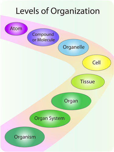
The human machine is organized at different levels, starting with the cell and ending with the entire organism (see Figure below). At each higher level of organization, there is a greater degree of complexity.

The human organism has several levels of organization.
Cells
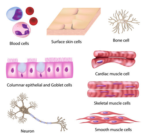
The most basic parts of the human machine are cells—an amazing 100 trillion of them by the time the average person reaches adulthood! Cells are the basic units of structure and function in the human body, as they are in all living things. Each cell carries out basic life processes that allow the body to survive. Many human cells are specialized in form and function, as shown in Figure below. Each type of cell in the figure plays a specific role. For example, nerve cells have long projections that help them carry electrical messages to other cells. Muscle cells have many mitochondria that provide the energy they need to move the body.

Different types of cells in the human body are specialized for specific jobs. Do you know the functions of any of the cell types shown here?
Tissues
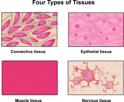
After the cell, the tissue is the next level of organization in the human body. A tissue is a group of connected cells that have a similar function. There are four basic types of human tissues: epithelial, muscle, nervous, and connective tissues. These four tissue types, which are shown in Figure below, make up all the organs of the human body.

The human body consists of these four tissue types.
- Connective tissue is made up of cells that form the body’s structure. Examples include bone and cartilage.
- Epithelial tissue is made up of cells that line inner and outer body surfaces, such as the skin and the lining of the digestive tract. Epithelial tissue protects the body and its internal organs, secretes substances such as hormones, and absorbs substances such as nutrients.
- Muscle tissue is made up of cells that have the unique ability to contract, or become shorter. Muscles attached to bones enable the body to move.
- Nervous tissue is made up of neurons, or nerve cells, that carry electrical messages. Nervous tissue makes up the brain and the nerves that connect the brain to all parts of the body.
Organs and Organ Systems
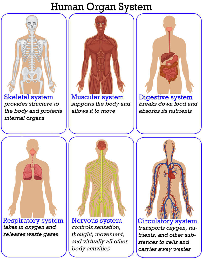
After tissues, organs are the next level of organization of the human body. An organ is a structure that consists of two or more types of tissues that work together to do the same job. Examples of human organs include the brain, heart, lungs, skin, and kidneys. Human organs are organized into organ systems, many of which are shown in Figure below. An organ system is a group of organs that work together to carry out a complex overall function. Each organ of the system does part of the larger job.
 Many of the organ systems that make up the human body are represented here. What is the overall function of each organ system?
Your body’s 12 organ systems are shown below (Table below). Your organ systems do not work alone in your body. They must all be able to work together. For example, one of the most important functions of organ systems is to provide cells with oxygen and nutrients and to remove toxic waste products such as carbon dioxide. A number of organ systems, including the cardiovascular and respiratory systems, all work together to do this.
| Organ System | Major Tissues and Organs | Function |
|---|---|---|
| Cardiovascular | Heart; blood vessels; blood | Transports oxygen, hormones, and nutrients to the body cells. Moves wastes and carbon dioxide away from cells. |
| Lymphatic | Lymph nodes; lymph vessels | Defend against infection and disease, moves lymph between tissues and the blood stream. |
| Digestive | Esophagus; stomach; small intestine; large intestine | Digests foods and absorbs nutrients, minerals, vitamins, and water. |
| Endocrine | Pituitary gland, hypothalamus; adrenal glands; ovaries; testes | Produces hormones that communicate between cells. |
| Integumentary | Skin, hair, nails | Provides protection from injury and water loss, physical defense against infection by microorganisms, and temperature control. |
| Muscular | Cardiac (heart) muscle; skeletal muscle; smooth muscle; tendons | Involved in movement and heat production. |
| Nervous | Brain, spinal cord; nerves | Collects, transfers, and processes information. |
| Reproductive |
Female: uterus; vagina; fallopian tubes; ovaries Male: penis; testes; seminal vesicles |
Produces gametes (sex cells) and sex hormones. |
| Respiratory | Trachea, larynx, pharynx, lungs | Brings air to sites where gas exchange can occur between the blood and cells (around body) or blood and air (lungs). |
| Skeletal | Bones, cartilage; ligaments | Supports and protects soft tissues of body; produces blood cells; stores minerals. |
| Urinary | Kidneys; urinary bladder | Removes extra water, salts, and waste products from blood and body; controls pH; controls water and salt balance. |
| Immune | Bone marrow; spleen; white blood cells | Defends against diseases. |
Summary
- The human body is organized at different levels, starting with the cell.
- Cells are organized into tissues, and tissues form organs.
- Organs are organized into organ systems such as the skeletal and muscular systems.
Review
- What are the levels of organization of the human body?
- Which type of tissue covers the surface of the body?
- What are the functions of the skeletal system?
- Which organ system supports the body and allows it to move?
- Explain how form and function are related in human cells. Include examples.
- Compare and contrast epithelial and muscle tissues.
Resources
| Image | Reference | Attributions |
 |
[Figure 1] | Credit: Rupali Raju Source: CK-12 Foundation License: CC BY-NC 3.0 |
 |
[Figure 2] | Credit: Image copyright Alila Medical Media, 2014;Image copyright Matthew Cole, 2014 Source: http://www.shutterstock.com License: Used under license from Shutterstock.com |
 |
[Figure 3] | Credit: Zachary Wilson Source: CK-12 Foundation License: CC BY-NC 3.0 |
 |
[Figure 4] | Credit: Image copyright Matthew Cole, 2014 Source: http://www.shutterstock.com License: Used under license from Shutterstock.com |

