
2.38: Cell Structure
- Page ID
- 8353
\( \newcommand{\vecs}[1]{\overset { \scriptstyle \rightharpoonup} {\mathbf{#1}} } \)
\( \newcommand{\vecd}[1]{\overset{-\!-\!\rightharpoonup}{\vphantom{a}\smash {#1}}} \)
\( \newcommand{\dsum}{\displaystyle\sum\limits} \)
\( \newcommand{\dint}{\displaystyle\int\limits} \)
\( \newcommand{\dlim}{\displaystyle\lim\limits} \)
\( \newcommand{\id}{\mathrm{id}}\) \( \newcommand{\Span}{\mathrm{span}}\)
( \newcommand{\kernel}{\mathrm{null}\,}\) \( \newcommand{\range}{\mathrm{range}\,}\)
\( \newcommand{\RealPart}{\mathrm{Re}}\) \( \newcommand{\ImaginaryPart}{\mathrm{Im}}\)
\( \newcommand{\Argument}{\mathrm{Arg}}\) \( \newcommand{\norm}[1]{\| #1 \|}\)
\( \newcommand{\inner}[2]{\langle #1, #2 \rangle}\)
\( \newcommand{\Span}{\mathrm{span}}\)
\( \newcommand{\id}{\mathrm{id}}\)
\( \newcommand{\Span}{\mathrm{span}}\)
\( \newcommand{\kernel}{\mathrm{null}\,}\)
\( \newcommand{\range}{\mathrm{range}\,}\)
\( \newcommand{\RealPart}{\mathrm{Re}}\)
\( \newcommand{\ImaginaryPart}{\mathrm{Im}}\)
\( \newcommand{\Argument}{\mathrm{Arg}}\)
\( \newcommand{\norm}[1]{\| #1 \|}\)
\( \newcommand{\inner}[2]{\langle #1, #2 \rangle}\)
\( \newcommand{\Span}{\mathrm{span}}\) \( \newcommand{\AA}{\unicode[.8,0]{x212B}}\)
\( \newcommand{\vectorA}[1]{\vec{#1}} % arrow\)
\( \newcommand{\vectorAt}[1]{\vec{\text{#1}}} % arrow\)
\( \newcommand{\vectorB}[1]{\overset { \scriptstyle \rightharpoonup} {\mathbf{#1}} } \)
\( \newcommand{\vectorC}[1]{\textbf{#1}} \)
\( \newcommand{\vectorD}[1]{\overrightarrow{#1}} \)
\( \newcommand{\vectorDt}[1]{\overrightarrow{\text{#1}}} \)
\( \newcommand{\vectE}[1]{\overset{-\!-\!\rightharpoonup}{\vphantom{a}\smash{\mathbf {#1}}}} \)
\( \newcommand{\vecs}[1]{\overset { \scriptstyle \rightharpoonup} {\mathbf{#1}} } \)
\(\newcommand{\longvect}{\overrightarrow}\)
\( \newcommand{\vecd}[1]{\overset{-\!-\!\rightharpoonup}{\vphantom{a}\smash {#1}}} \)
\(\newcommand{\avec}{\mathbf a}\) \(\newcommand{\bvec}{\mathbf b}\) \(\newcommand{\cvec}{\mathbf c}\) \(\newcommand{\dvec}{\mathbf d}\) \(\newcommand{\dtil}{\widetilde{\mathbf d}}\) \(\newcommand{\evec}{\mathbf e}\) \(\newcommand{\fvec}{\mathbf f}\) \(\newcommand{\nvec}{\mathbf n}\) \(\newcommand{\pvec}{\mathbf p}\) \(\newcommand{\qvec}{\mathbf q}\) \(\newcommand{\svec}{\mathbf s}\) \(\newcommand{\tvec}{\mathbf t}\) \(\newcommand{\uvec}{\mathbf u}\) \(\newcommand{\vvec}{\mathbf v}\) \(\newcommand{\wvec}{\mathbf w}\) \(\newcommand{\xvec}{\mathbf x}\) \(\newcommand{\yvec}{\mathbf y}\) \(\newcommand{\zvec}{\mathbf z}\) \(\newcommand{\rvec}{\mathbf r}\) \(\newcommand{\mvec}{\mathbf m}\) \(\newcommand{\zerovec}{\mathbf 0}\) \(\newcommand{\onevec}{\mathbf 1}\) \(\newcommand{\real}{\mathbb R}\) \(\newcommand{\twovec}[2]{\left[\begin{array}{r}#1 \\ #2 \end{array}\right]}\) \(\newcommand{\ctwovec}[2]{\left[\begin{array}{c}#1 \\ #2 \end{array}\right]}\) \(\newcommand{\threevec}[3]{\left[\begin{array}{r}#1 \\ #2 \\ #3 \end{array}\right]}\) \(\newcommand{\cthreevec}[3]{\left[\begin{array}{c}#1 \\ #2 \\ #3 \end{array}\right]}\) \(\newcommand{\fourvec}[4]{\left[\begin{array}{r}#1 \\ #2 \\ #3 \\ #4 \end{array}\right]}\) \(\newcommand{\cfourvec}[4]{\left[\begin{array}{c}#1 \\ #2 \\ #3 \\ #4 \end{array}\right]}\) \(\newcommand{\fivevec}[5]{\left[\begin{array}{r}#1 \\ #2 \\ #3 \\ #4 \\ #5 \\ \end{array}\right]}\) \(\newcommand{\cfivevec}[5]{\left[\begin{array}{c}#1 \\ #2 \\ #3 \\ #4 \\ #5 \\ \end{array}\right]}\) \(\newcommand{\mattwo}[4]{\left[\begin{array}{rr}#1 \amp #2 \\ #3 \amp #4 \\ \end{array}\right]}\) \(\newcommand{\laspan}[1]{\text{Span}\{#1\}}\) \(\newcommand{\bcal}{\cal B}\) \(\newcommand{\ccal}{\cal C}\) \(\newcommand{\scal}{\cal S}\) \(\newcommand{\wcal}{\cal W}\) \(\newcommand{\ecal}{\cal E}\) \(\newcommand{\coords}[2]{\left\{#1\right\}_{#2}}\) \(\newcommand{\gray}[1]{\color{gray}{#1}}\) \(\newcommand{\lgray}[1]{\color{lightgray}{#1}}\) \(\newcommand{\rank}{\operatorname{rank}}\) \(\newcommand{\row}{\text{Row}}\) \(\newcommand{\col}{\text{Col}}\) \(\renewcommand{\row}{\text{Row}}\) \(\newcommand{\nul}{\text{Nul}}\) \(\newcommand{\var}{\text{Var}}\) \(\newcommand{\corr}{\text{corr}}\) \(\newcommand{\len}[1]{\left|#1\right|}\) \(\newcommand{\bbar}{\overline{\bvec}}\) \(\newcommand{\bhat}{\widehat{\bvec}}\) \(\newcommand{\bperp}{\bvec^\perp}\) \(\newcommand{\xhat}{\widehat{\xvec}}\) \(\newcommand{\vhat}{\widehat{\vvec}}\) \(\newcommand{\uhat}{\widehat{\uvec}}\) \(\newcommand{\what}{\widehat{\wvec}}\) \(\newcommand{\Sighat}{\widehat{\Sigma}}\) \(\newcommand{\lt}{<}\) \(\newcommand{\gt}{>}\) \(\newcommand{\amp}{&}\) \(\definecolor{fillinmathshade}{gray}{0.9}\)
Does a cell have its own ER?
Yes, but in this case, the ER is not just for emergencies. True, there might be times when the cell responds to emergency conditions and the functions of the ER may be needed, but usually the cell's ER is involved in normal functions. Proteins are also made on the outside of the ER, and this starts a whole process of protein transport, both around the inside of the cell and to the cell membrane and out.
Other Organelles
In addition to the nucleus, eukaryotic cells have many other organelles, including the endoplasmic reticulum, Golgi apparatus, vesicles, vacuoles, and centrioles.
Endoplasmic Reticulum
The endoplasmic reticulum (ER) (plural, reticuli) is a network of phospholipid membranes that form hollow tubes, flattened sheets, and round sacs. These flattened, hollow folds and sacs are called cisternae. The ER has two major functions:
- Transport: Molecules, such as proteins, can move from place to place inside the ER, much like on an intracellular highway.
- Synthesis: Ribosomes that are attached to ER, similar to unattached ribosomes, make proteins. Lipids are also produced in the ER.
There are two types of endoplasmic reticulum, rough endoplasmic reticulum (RER) and smooth endoplasmic reticulum (SER).
- Rough endoplasmic reticulum is studded with ribosomes, which gives it a "rough" appearance. These ribosomes make proteins that are then transported from the ER in small sacs called transport vesicles. The transport vesicles pinch off the ends of the ER. The rough endoplasmic reticulum works with the Golgi apparatus to move new proteins to their proper destinations in the cell. The membrane of the RER is continuous with the outer layer of the nuclear envelope.
- Smooth endoplasmic reticulum does not have any ribosomes attached to it, and so it has a smooth appearance. SER has many different functions, some of which include lipid synthesis, calcium ion storage, and drug detoxification. Smooth endoplasmic reticulum is found in both animal and plant cells and it serves different functions in each. The SER is made up of tubules and vesicles that branch out to form a network. In some cells there are dilated areas like the sacs of RER. Smooth endoplasmic reticulum and RER form an interconnected network.
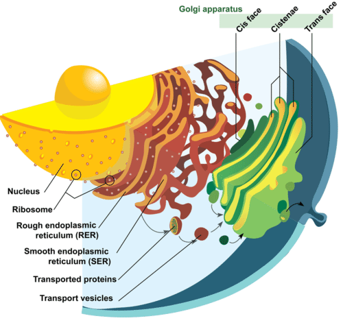 Image of nucleus, endoplasmic reticulum and Golgi apparatus, and how they work together. The process of secretion from endoplasmic reticuli to Golgi apparatus is shown.
Golgi Apparatus
The Golgi apparatus is a large organelle that is usually made up of five to eight cup-shaped, membrane-covered discs called cisternae, as shown in the Figure above. The cisternae look a bit like a stack of deflated balloons. The Golgi apparatus modifies, sorts, and packages different substances for secretion out of the cell, or for use within the cell. The Golgi apparatus is found close to the nucleus of the cell, where it modifies proteins that have been delivered in transport vesicles from the RER. It is also involved in the transport of lipids around the cell. Pieces of the Golgi membrane pinch off to form vesicles that transport molecules around the cell. The Golgi apparatus can be thought of as similar to a post office; it packages and labels "items" and then sends them to different parts of the cell. Both plant and animal cells have a Golgi apparatus. Plant cells can have up to several hundred Golgi stacks scattered throughout the cytoplasm. In plants, the Golgi apparatus contains enzymes that synthesize some of the cell wall polysaccharides.
Vesicles
A vesicle is a small, spherical compartment that is separated from the cytosol by at least one lipid bilayer. Many vesicles are made in the Golgi apparatus and the endoplasmic reticulum, or are made from parts of the cell membrane. Vesicles from the Golgi apparatus can be seen in the Figure above. Because it is separated from the cytosol, the space inside the vesicle can be made to be chemically different from the cytosol. Vesicles are basic tools of the cell for organizing metabolism, transport, and storage of molecules. Vesicles are also used as chemical reaction chambers. They can be classified by their contents and function.
- Transport vesicles are able to move molecules between locations inside the cell. For example, transport vesicles move proteins from the rough endoplasmic reticulum to the Golgi apparatus.
- Lysosomes are vesicles that are formed by the Golgi apparatus. They contain powerful enzymes that could break down (digest) the cell. Lysosomes break down harmful cell products, waste materials, and cellular debris and then force them out of the cell. They also digest invading organisms such as bacteria. Lysosomes also break down cells that are ready to die, a process called autolysis.
- Peroxisomes are vesicles that use oxygen to break down toxic substances in the cell. Unlike lysosomes, which are formed by the Golgi apparatus, peroxisomes self-replicate by growing bigger and then dividing. They are common in liver and kidney cells that break down harmful substances. Peroxisomes are named for the hydrogen peroxide (H2O2) that is produced when they break down organic compounds. Hydrogen peroxide is toxic, and in turn is broken down into water (H2O) and oxygen (O2) molecules.
Vacuoles
Vacuoles are membrane-bound organelles that can have secretory, excretory, and storage functions. Many organisms will use vacuoles as storage areas and some plant cells have very large vacuoles. Vesicles are much smaller than vacuoles and function in transporting materials both within and to the outside of the cell.
Centrioles
Centrioles are rod-like structures made of short microtubules. Nine groups of three microtubules make up each centriole. Two perpendicular centrioles make up the centrosome. Centrioles are very important in cellular division, where they arrange the mitotic spindles that pull the chromosome apart during mitosis.
Summary
- The endoplasmic reticulum (ER) is involved in the synthesis of lipids and synthesis and transport of proteins.
- The Golgi apparatus modifies, sorts, and packages different substances for secretion out of the cell, or for use within the cell.
- Vesicles are also used as chemical reaction chambers. Transport vesicles, lysosomes, and peroxisomes are types of vesicles.
- Vacuoles have secretory, excretory, and storage functions.
- Centrioles are made of short microtubules and are very important in cell division.
Review
- List five organelles eukaryotes have that prokaryotes do not have.
- Explain how the following organelles ensure that a cell has the proteins it needs: nucleus, rough ER, vesicles, and Golgi apparatus.
- What is the main difference between rough endoplasmic reticulum and smooth endoplasmic reticulum?
- Describe the three types of vesicles.
| Image | Reference | Attributions |
 |
[Figure 1] | Source: (a) commons.wikimedia.org/wiki/File:FluorescentCells.jpg ; (b) commons.wikimedia.org/wiki/File:E7_amnion_cells.png License: CC BY-NC |
 |
[Figure 2] | Credit: Mariana Ruiz Villarreal (LadyofHats), modified for CK-12 Foundation Source: Original version: commons.wikimedia.org/wiki/File:Endomembrane_system_diagram_en.svg ; (a) commons.wikimedia.org/wiki/File:FluorescentCells.jpg ; (b) commons.wikimedia.org/wiki/File:E7_amnion_cells.png License: Public Domain |