
2.37: Ribosomes and Mitochondria
- Page ID
- 8352
\( \newcommand{\vecs}[1]{\overset { \scriptstyle \rightharpoonup} {\mathbf{#1}} } \)
\( \newcommand{\vecd}[1]{\overset{-\!-\!\rightharpoonup}{\vphantom{a}\smash {#1}}} \)
\( \newcommand{\dsum}{\displaystyle\sum\limits} \)
\( \newcommand{\dint}{\displaystyle\int\limits} \)
\( \newcommand{\dlim}{\displaystyle\lim\limits} \)
\( \newcommand{\id}{\mathrm{id}}\) \( \newcommand{\Span}{\mathrm{span}}\)
( \newcommand{\kernel}{\mathrm{null}\,}\) \( \newcommand{\range}{\mathrm{range}\,}\)
\( \newcommand{\RealPart}{\mathrm{Re}}\) \( \newcommand{\ImaginaryPart}{\mathrm{Im}}\)
\( \newcommand{\Argument}{\mathrm{Arg}}\) \( \newcommand{\norm}[1]{\| #1 \|}\)
\( \newcommand{\inner}[2]{\langle #1, #2 \rangle}\)
\( \newcommand{\Span}{\mathrm{span}}\)
\( \newcommand{\id}{\mathrm{id}}\)
\( \newcommand{\Span}{\mathrm{span}}\)
\( \newcommand{\kernel}{\mathrm{null}\,}\)
\( \newcommand{\range}{\mathrm{range}\,}\)
\( \newcommand{\RealPart}{\mathrm{Re}}\)
\( \newcommand{\ImaginaryPart}{\mathrm{Im}}\)
\( \newcommand{\Argument}{\mathrm{Arg}}\)
\( \newcommand{\norm}[1]{\| #1 \|}\)
\( \newcommand{\inner}[2]{\langle #1, #2 \rangle}\)
\( \newcommand{\Span}{\mathrm{span}}\) \( \newcommand{\AA}{\unicode[.8,0]{x212B}}\)
\( \newcommand{\vectorA}[1]{\vec{#1}} % arrow\)
\( \newcommand{\vectorAt}[1]{\vec{\text{#1}}} % arrow\)
\( \newcommand{\vectorB}[1]{\overset { \scriptstyle \rightharpoonup} {\mathbf{#1}} } \)
\( \newcommand{\vectorC}[1]{\textbf{#1}} \)
\( \newcommand{\vectorD}[1]{\overrightarrow{#1}} \)
\( \newcommand{\vectorDt}[1]{\overrightarrow{\text{#1}}} \)
\( \newcommand{\vectE}[1]{\overset{-\!-\!\rightharpoonup}{\vphantom{a}\smash{\mathbf {#1}}}} \)
\( \newcommand{\vecs}[1]{\overset { \scriptstyle \rightharpoonup} {\mathbf{#1}} } \)
\(\newcommand{\longvect}{\overrightarrow}\)
\( \newcommand{\vecd}[1]{\overset{-\!-\!\rightharpoonup}{\vphantom{a}\smash {#1}}} \)
\(\newcommand{\avec}{\mathbf a}\) \(\newcommand{\bvec}{\mathbf b}\) \(\newcommand{\cvec}{\mathbf c}\) \(\newcommand{\dvec}{\mathbf d}\) \(\newcommand{\dtil}{\widetilde{\mathbf d}}\) \(\newcommand{\evec}{\mathbf e}\) \(\newcommand{\fvec}{\mathbf f}\) \(\newcommand{\nvec}{\mathbf n}\) \(\newcommand{\pvec}{\mathbf p}\) \(\newcommand{\qvec}{\mathbf q}\) \(\newcommand{\svec}{\mathbf s}\) \(\newcommand{\tvec}{\mathbf t}\) \(\newcommand{\uvec}{\mathbf u}\) \(\newcommand{\vvec}{\mathbf v}\) \(\newcommand{\wvec}{\mathbf w}\) \(\newcommand{\xvec}{\mathbf x}\) \(\newcommand{\yvec}{\mathbf y}\) \(\newcommand{\zvec}{\mathbf z}\) \(\newcommand{\rvec}{\mathbf r}\) \(\newcommand{\mvec}{\mathbf m}\) \(\newcommand{\zerovec}{\mathbf 0}\) \(\newcommand{\onevec}{\mathbf 1}\) \(\newcommand{\real}{\mathbb R}\) \(\newcommand{\twovec}[2]{\left[\begin{array}{r}#1 \\ #2 \end{array}\right]}\) \(\newcommand{\ctwovec}[2]{\left[\begin{array}{c}#1 \\ #2 \end{array}\right]}\) \(\newcommand{\threevec}[3]{\left[\begin{array}{r}#1 \\ #2 \\ #3 \end{array}\right]}\) \(\newcommand{\cthreevec}[3]{\left[\begin{array}{c}#1 \\ #2 \\ #3 \end{array}\right]}\) \(\newcommand{\fourvec}[4]{\left[\begin{array}{r}#1 \\ #2 \\ #3 \\ #4 \end{array}\right]}\) \(\newcommand{\cfourvec}[4]{\left[\begin{array}{c}#1 \\ #2 \\ #3 \\ #4 \end{array}\right]}\) \(\newcommand{\fivevec}[5]{\left[\begin{array}{r}#1 \\ #2 \\ #3 \\ #4 \\ #5 \\ \end{array}\right]}\) \(\newcommand{\cfivevec}[5]{\left[\begin{array}{c}#1 \\ #2 \\ #3 \\ #4 \\ #5 \\ \end{array}\right]}\) \(\newcommand{\mattwo}[4]{\left[\begin{array}{rr}#1 \amp #2 \\ #3 \amp #4 \\ \end{array}\right]}\) \(\newcommand{\laspan}[1]{\text{Span}\{#1\}}\) \(\newcommand{\bcal}{\cal B}\) \(\newcommand{\ccal}{\cal C}\) \(\newcommand{\scal}{\cal S}\) \(\newcommand{\wcal}{\cal W}\) \(\newcommand{\ecal}{\cal E}\) \(\newcommand{\coords}[2]{\left\{#1\right\}_{#2}}\) \(\newcommand{\gray}[1]{\color{gray}{#1}}\) \(\newcommand{\lgray}[1]{\color{lightgray}{#1}}\) \(\newcommand{\rank}{\operatorname{rank}}\) \(\newcommand{\row}{\text{Row}}\) \(\newcommand{\col}{\text{Col}}\) \(\renewcommand{\row}{\text{Row}}\) \(\newcommand{\nul}{\text{Nul}}\) \(\newcommand{\var}{\text{Var}}\) \(\newcommand{\corr}{\text{corr}}\) \(\newcommand{\len}[1]{\left|#1\right|}\) \(\newcommand{\bbar}{\overline{\bvec}}\) \(\newcommand{\bhat}{\widehat{\bvec}}\) \(\newcommand{\bperp}{\bvec^\perp}\) \(\newcommand{\xhat}{\widehat{\xvec}}\) \(\newcommand{\vhat}{\widehat{\vvec}}\) \(\newcommand{\uhat}{\widehat{\uvec}}\) \(\newcommand{\what}{\widehat{\wvec}}\) \(\newcommand{\Sighat}{\widehat{\Sigma}}\) \(\newcommand{\lt}{<}\) \(\newcommand{\gt}{>}\) \(\newcommand{\amp}{&}\) \(\definecolor{fillinmathshade}{gray}{0.9}\)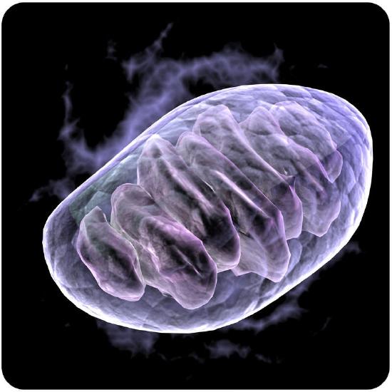
Sperm cells and muscle cells need lots of energy. What do they have in common?
They have lots of mitochondria. Mitochondria are called the power plants of the cell, as these organelles are where most of the cell's energy is produced. Cells that need lots of energy have lots of mitochondria.
Other Organelles
In addition to the nucleus, eukaryotic cells have many other organelles, including ribosomes and mitochondria. Ribosomes are present in all cells.
Ribosomes
Ribosomes are small organelles and are the sites of protein synthesis (or assembly). They are made of ribosomal protein and ribosomal RNA, and are found in both eukaryotic and prokaryotic cells. Unlike other organelles, ribosomes are not surrounded by a membrane. Each ribosome has two parts, a large and a small subunit, as shown in the Figure below. The subunits are attached to one another. Ribosomes can be found alone or in groups within the cytoplasm. Some ribosomes are attached to the endoplasmic reticulum (ER), and others are attached to the nuclear envelope.
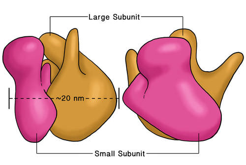 The two subunits that make up a ribosome, small organelles that are intercellular protein factories.
Ribozymes are RNA molecules that catalyze chemical reactions, such as translation. Translation is the process of ordering the amino acids in the assembly of a protein, and translation will be discussed more in another concept. Briefly, the ribosomes interact with other RNA molecules to make chains of amino acids called polypeptide chains, due to the peptide bond that forms between individual amino acids. Polypeptide chains are built from the genetic instructions held within a messenger RNA (mRNA) molecule. Polypeptide chains that are made on the rough ER are inserted directly into the ER and then are transported to their various cellular destinations. Ribosomes on the rough ER usually produce proteins that are destined for the cell membrane.
Ribosomes are found in both eukaryotic and prokaryotic cells. Ribosomes are not surrounded by a membrane. The other organelles found in eukaryotic cells are surrounded by a membrane.
Mitochondria
A mitochondrion (mitochondria, plural), is a membrane-enclosed organelle that is found in most eukaryotic cells. Mitochondria are called the "power plants" of the cell because they are the sites of cellular respiration, where they use energy from organic compounds to make ATP (adenosine triphosphate). ATP is the cell's energy source that is used for such things such as movement and cell division. Some ATP is made in the cytosol of the cell, but most of it is made inside mitochondria. The number of mitochondria in a cell depends on the cell’s energy needs. For example, active human muscle cells may have thousands of mitochondria, while less active red blood cells do not have any.
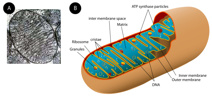
As the Figure above (a) and (b) show, a mitochondrion has two phospholipid membranes. The smooth outer membrane separates the mitochondrion from the cytosol. The inner membrane has many folds, called cristae. The fluid-filled inside of the mitochondrion, called the matrix, is where most of the cell’s ATP is made.
Although most of a cell's DNA is contained in the cell nucleus, mitochondria have their own DNA. Mitochondria are able to reproduce asexually, and scientists think that they are descended from prokaryotes. According to the endosymbiotic theory, mitochondria were once free-living prokaryotes that infected or were engulfed by ancient eukaryotic cells. The invading prokaryotes were protected inside the eukaryotic host cell, and in turn the prokaryote supplied extra ATP to its host.
Summary
- Ribosomes are small organelles and are the site of protein synthesis. Ribosomes are found in all cells.
- Mitochondria are where energy from organic compounds is used to make ATP.
Review
- What is the function of a ribosome?
- What is a significant difference between the structure of a ribosome and other organelles?
- Identify the reason why mitochondria are called "power plants" of the cell.
- Describe the structure of a mitochondrion.
| Image | Reference | Attributions |
 |
[Figure 1] | Source: (a) commons.wikimedia.org/wiki/File:FluorescentCells.jpg ; (b) commons.wikimedia.org/wiki/File:E7_amnion_cells.png License: CC BY-NC |
 |
[Figure 2] | Credit: Laura Guerin;Mariana Ruiz Villarreal (LadyofHats), modified for CK-12 Foundation;(a) Courtesy of the National Institutes of Health; (b) Mariana Ruiz Villarreal (User:LadyofHats/Wikimedia Commons) Source: CK-12 Foundation ; Original version: commons.wikimedia.org/wiki/File:Endomembrane_system_diagram_en.svg ; (a) en.Wikipedia.org/wiki/File:Mitochondrion_186.jpg ; (b) commons.wikimedia.org/wiki/File:Animal_mitochondrion_diagram_en.svg ; (a) commons.wikimedia.org/wiki/File:FluorescentCells.jpg ; (b) commons.wikimedia.org/wiki/File:E7_amnion_cells.png License: CC BY-NC 3.0; Public Domain |
|
[Figure 3] | Credit: (a) Courtesy of the National Institutes of Health; (b) Mariana Ruiz Villarreal (User:LadyofHats/Wikimedia Commons);Mariana Ruiz Villarreal (LadyofHats), modified for CK-12 Foundation Source: (a) en.Wikipedia.org/wiki/File:Mitochondrion_186.jpg ; (b) commons.wikimedia.org/wiki/File:Animal_mitochondrion_diagram_en.svg ; Original version: commons.wikimedia.org/wiki/File:Endomembrane_system_diagram_en.svg ; (a) commons.wikimedia.org/wiki/File:FluorescentCells.jpg ; (b) commons.wikimedia.org/wiki/File:E7_amnion_cells.png License: Public Domain |