
7.11: Bacteria Structure
- Page ID
- 1461
\( \newcommand{\vecs}[1]{\overset { \scriptstyle \rightharpoonup} {\mathbf{#1}} } \)
\( \newcommand{\vecd}[1]{\overset{-\!-\!\rightharpoonup}{\vphantom{a}\smash {#1}}} \)
\( \newcommand{\dsum}{\displaystyle\sum\limits} \)
\( \newcommand{\dint}{\displaystyle\int\limits} \)
\( \newcommand{\dlim}{\displaystyle\lim\limits} \)
\( \newcommand{\id}{\mathrm{id}}\) \( \newcommand{\Span}{\mathrm{span}}\)
( \newcommand{\kernel}{\mathrm{null}\,}\) \( \newcommand{\range}{\mathrm{range}\,}\)
\( \newcommand{\RealPart}{\mathrm{Re}}\) \( \newcommand{\ImaginaryPart}{\mathrm{Im}}\)
\( \newcommand{\Argument}{\mathrm{Arg}}\) \( \newcommand{\norm}[1]{\| #1 \|}\)
\( \newcommand{\inner}[2]{\langle #1, #2 \rangle}\)
\( \newcommand{\Span}{\mathrm{span}}\)
\( \newcommand{\id}{\mathrm{id}}\)
\( \newcommand{\Span}{\mathrm{span}}\)
\( \newcommand{\kernel}{\mathrm{null}\,}\)
\( \newcommand{\range}{\mathrm{range}\,}\)
\( \newcommand{\RealPart}{\mathrm{Re}}\)
\( \newcommand{\ImaginaryPart}{\mathrm{Im}}\)
\( \newcommand{\Argument}{\mathrm{Arg}}\)
\( \newcommand{\norm}[1]{\| #1 \|}\)
\( \newcommand{\inner}[2]{\langle #1, #2 \rangle}\)
\( \newcommand{\Span}{\mathrm{span}}\) \( \newcommand{\AA}{\unicode[.8,0]{x212B}}\)
\( \newcommand{\vectorA}[1]{\vec{#1}} % arrow\)
\( \newcommand{\vectorAt}[1]{\vec{\text{#1}}} % arrow\)
\( \newcommand{\vectorB}[1]{\overset { \scriptstyle \rightharpoonup} {\mathbf{#1}} } \)
\( \newcommand{\vectorC}[1]{\textbf{#1}} \)
\( \newcommand{\vectorD}[1]{\overrightarrow{#1}} \)
\( \newcommand{\vectorDt}[1]{\overrightarrow{\text{#1}}} \)
\( \newcommand{\vectE}[1]{\overset{-\!-\!\rightharpoonup}{\vphantom{a}\smash{\mathbf {#1}}}} \)
\( \newcommand{\vecs}[1]{\overset { \scriptstyle \rightharpoonup} {\mathbf{#1}} } \)
\(\newcommand{\longvect}{\overrightarrow}\)
\( \newcommand{\vecd}[1]{\overset{-\!-\!\rightharpoonup}{\vphantom{a}\smash {#1}}} \)
\(\newcommand{\avec}{\mathbf a}\) \(\newcommand{\bvec}{\mathbf b}\) \(\newcommand{\cvec}{\mathbf c}\) \(\newcommand{\dvec}{\mathbf d}\) \(\newcommand{\dtil}{\widetilde{\mathbf d}}\) \(\newcommand{\evec}{\mathbf e}\) \(\newcommand{\fvec}{\mathbf f}\) \(\newcommand{\nvec}{\mathbf n}\) \(\newcommand{\pvec}{\mathbf p}\) \(\newcommand{\qvec}{\mathbf q}\) \(\newcommand{\svec}{\mathbf s}\) \(\newcommand{\tvec}{\mathbf t}\) \(\newcommand{\uvec}{\mathbf u}\) \(\newcommand{\vvec}{\mathbf v}\) \(\newcommand{\wvec}{\mathbf w}\) \(\newcommand{\xvec}{\mathbf x}\) \(\newcommand{\yvec}{\mathbf y}\) \(\newcommand{\zvec}{\mathbf z}\) \(\newcommand{\rvec}{\mathbf r}\) \(\newcommand{\mvec}{\mathbf m}\) \(\newcommand{\zerovec}{\mathbf 0}\) \(\newcommand{\onevec}{\mathbf 1}\) \(\newcommand{\real}{\mathbb R}\) \(\newcommand{\twovec}[2]{\left[\begin{array}{r}#1 \\ #2 \end{array}\right]}\) \(\newcommand{\ctwovec}[2]{\left[\begin{array}{c}#1 \\ #2 \end{array}\right]}\) \(\newcommand{\threevec}[3]{\left[\begin{array}{r}#1 \\ #2 \\ #3 \end{array}\right]}\) \(\newcommand{\cthreevec}[3]{\left[\begin{array}{c}#1 \\ #2 \\ #3 \end{array}\right]}\) \(\newcommand{\fourvec}[4]{\left[\begin{array}{r}#1 \\ #2 \\ #3 \\ #4 \end{array}\right]}\) \(\newcommand{\cfourvec}[4]{\left[\begin{array}{c}#1 \\ #2 \\ #3 \\ #4 \end{array}\right]}\) \(\newcommand{\fivevec}[5]{\left[\begin{array}{r}#1 \\ #2 \\ #3 \\ #4 \\ #5 \\ \end{array}\right]}\) \(\newcommand{\cfivevec}[5]{\left[\begin{array}{c}#1 \\ #2 \\ #3 \\ #4 \\ #5 \\ \end{array}\right]}\) \(\newcommand{\mattwo}[4]{\left[\begin{array}{rr}#1 \amp #2 \\ #3 \amp #4 \\ \end{array}\right]}\) \(\newcommand{\laspan}[1]{\text{Span}\{#1\}}\) \(\newcommand{\bcal}{\cal B}\) \(\newcommand{\ccal}{\cal C}\) \(\newcommand{\scal}{\cal S}\) \(\newcommand{\wcal}{\cal W}\) \(\newcommand{\ecal}{\cal E}\) \(\newcommand{\coords}[2]{\left\{#1\right\}_{#2}}\) \(\newcommand{\gray}[1]{\color{gray}{#1}}\) \(\newcommand{\lgray}[1]{\color{lightgray}{#1}}\) \(\newcommand{\rank}{\operatorname{rank}}\) \(\newcommand{\row}{\text{Row}}\) \(\newcommand{\col}{\text{Col}}\) \(\renewcommand{\row}{\text{Row}}\) \(\newcommand{\nul}{\text{Nul}}\) \(\newcommand{\var}{\text{Var}}\) \(\newcommand{\corr}{\text{corr}}\) \(\newcommand{\len}[1]{\left|#1\right|}\) \(\newcommand{\bbar}{\overline{\bvec}}\) \(\newcommand{\bhat}{\widehat{\bvec}}\) \(\newcommand{\bperp}{\bvec^\perp}\) \(\newcommand{\xhat}{\widehat{\xvec}}\) \(\newcommand{\vhat}{\widehat{\vvec}}\) \(\newcommand{\uhat}{\widehat{\uvec}}\) \(\newcommand{\what}{\widehat{\wvec}}\) \(\newcommand{\Sighat}{\widehat{\Sigma}}\) \(\newcommand{\lt}{<}\) \(\newcommand{\gt}{>}\) \(\newcommand{\amp}{&}\) \(\definecolor{fillinmathshade}{gray}{0.9}\)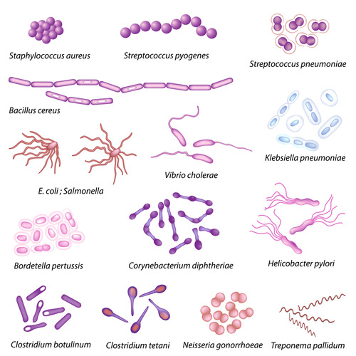
Does the shape matter?
It does if you're a bacterium. Prokaryotic cells are distinguished by their shape. And as you can imagine, shape may have something to do with mobility.
Prokaryote Structure
Most prokaryotic cells are much smaller than eukaryotic cells. Although they are tiny, prokaryotic cells can be distinguished by their shapes. The most common shapes are helices, spheres, and rods (see Figure below).
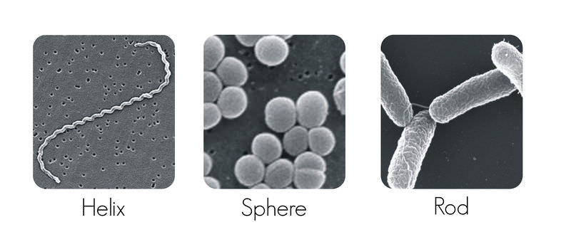 Prokaryotic Cell Shapes. The three most common prokaryotic cell shapes are shown here.
Plasma Membrane and Cell Wall
Like other cells, prokaryotic cells have a plasma membrane (see Figure below). It controls what enters and leaves the cell. It is also the site of many metabolic reactions. For example, cellular respiration and photosynthesis take place in the plasma membrane.
Most prokaryotes also have a cell wall. It lies just outside the plasma membrane. It gives strength and rigidity to the cell. Bacteria and Archaea differ in the makeup of their cell wall. The cell wall of Bacteria contains peptidoglycan, composed of sugars and amino acids. The cell wall of most Archaea lacks peptidoglycan.
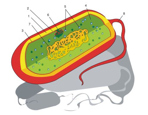 Prokaryotic Cell. The main parts of a prokaryotic cell are shown in this diagram. The structure called a mesosome was once thought to be an organelle. More evidence has convinced most scientists that it is not a true cell structure at all. Instead, it seems to be an artifact of cell preparation. This is a good example of how scientific knowledge is revised as more evidence becomes available. Can you identify each of the labeled structures?
Cytoplasm and Cell Structures
Inside the plasma membrane of prokaryotic cells is the cytoplasm. It contains several structures, including ribosomes, a cytoskeleton, and genetic material. Ribosomes are sites where proteins are made. The cytoskeleton helps the cell keep its shape. The genetic material is usually a single loop of DNA. There may also be small, circular pieces of DNA, called plasmids. (see Figure below). The cytoplasm may contain microcompartments as well. These are tiny structures enclosed by proteins. They contain enzymes and are involved in metabolic processes.
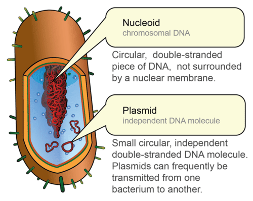
Prokaryotic DNA. The DNA of a prokaryotic cell is in the cytoplasm because the cell lacks a nucleus.
Extracellular Structures
Many prokaryotes have an extra layer, called a capsule, outside the cell wall. The capsule protects the cell from chemicals and from drying out. It also allows the cell to stick to surfaces and to other cells. Because of this, many prokaryotes can form biofilms, like the one shown in Figure below. A biofilm is a colony of prokaryotes that is stuck to a surface such as a rock or a host’s tissues. The sticky plaque that collects on your teeth between brushings is a biofilm. It consists of millions of bacteria.
Most prokaryotes also have long, thin protein structures called flagella (singular, flagellum). They extend from the plasma membrane. Flagella help prokaryotes move. They spin around a fixed base, causing the cell to roll and tumble. As shown in Figure below, prokaryotes may have one or more flagella.
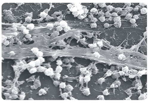 Bacterial Biofilm. The greatly magnified biofilm shown here was found on a medical catheter (tubing) removed from a patient’s body.
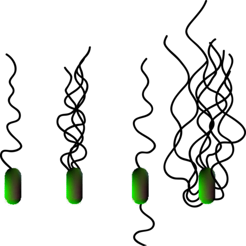 Variations in the Flagella of Bacteria. Flagella in prokaryotes may be located at one or both ends of the cell or all around it. They help prokaryotes move toward food or away from toxins.
Endospores
Many organisms form spores for reproduction. Some prokaryotes form spores for survival. Called endospores, they form inside prokaryotic cells when they are under stress. The stress could be UV radiation, high temperatures, or harsh chemicals. Endospores enclose the DNA and help it survive under conditions that may kill the cell. Endospores are commonly found in soil and water. They may survive for long periods of time.
Summary
- Most prokaryotic cells are much smaller than eukaryotic cells.
- Prokaryotic cells have a cell wall outside their plasma membrane.
- Prokaryotic DNA consists of a single loop. Some prokaryotes also have small, circular pieces of DNA called plasmids.
Review
- Identify the three most common shapes of prokaryotic cells.
- Describe a typical prokaryotic cell.
- What are the roles of flagella and endospores in prokaryotes?
| Image | Reference | Attributions |
|
[Figure 1] | Credit: Gram cell wall: User:JulianOnions/Wikimedia Commons; Gram-positive: JA Jernigan et al./Centers for Disease Control and Prevention; Gram-negative: William A. Clark/Centers for Disease and Control;Gram cell wall: commons.wikimedia.org/wiki/Fi...-Cell-Wall.jpg; Gram-positive: http://www.cdc.gov/ncidod/EID/vol7no6/jerniganG2.htm; Gram-negative: commons.wikimedia.org/wiki/Fi...agulans_01.jpg Source: Gram cell wall: commons.wikimedia.org/wiki/File:Gram-Cell-Wall.jpg ; Gram-positive: www.cdc.gov/ncidod/EID/vol7no6/jerniganG2.htm ; Gram-negative: commons.wikimedia.org/wiki/File:Bacillus_coagulans_01.jpg License: CC BY-NC |
|
[Figure 2] | Credit: Helix: Janice Carr/Centers for Disease Control and Prevention; Sphere: Janice Carr/Centers for Disease Control and Prevention; Rod: Volker Brinkmann, Max Planck Institute for Infection Biology, Berlin, Germany Source: Helix: commons.wikimedia.org/wiki/File:Leptospira_interrogans_strain_RGA_01.png ; Sphere: commons.wikimedia.org/wiki/File:Staphylococcus_aureus_01.jpg ; Rod: commons.wikimedia.org/wiki/File:Salmonella_typhimurium.png License: (Helix and Sphere) Public Domain; (Rod) CC BY 2.5 |
 |
[Figure 3] | Credit: Mariana Ruiz Villarreal (User:LadyofHats/Wikimedia Commons) Source: commons.wikimedia.org/wiki/File:Prokaryote_cell_diagram_international.svg License: Public Domain |
 |
[Figure 4] | Credit: Mariana Ruiz Villarreal (LadyofHats) for CK-12 Foundation Source: CK-12 Foundation License: CC BY-NC 3.0 |
 |
[Figure 5] | Credit: Courtesy of Janice Carr/Centers for Disease Control and Prevention Source: commons.wikimedia.org/wiki/File:Staphylococcus_on_catheter.png License: Public Domain |
 |
[Figure 6] | Credit: User:Adenosine/Wikimedia Commons Source: commons.wikimedia.org/wiki/File:Flagella.svg License: CC BY 3.0 |