
2.34: Membrane Proteins
- Page ID
- 1442
\( \newcommand{\vecs}[1]{\overset { \scriptstyle \rightharpoonup} {\mathbf{#1}} } \)
\( \newcommand{\vecd}[1]{\overset{-\!-\!\rightharpoonup}{\vphantom{a}\smash {#1}}} \)
\( \newcommand{\dsum}{\displaystyle\sum\limits} \)
\( \newcommand{\dint}{\displaystyle\int\limits} \)
\( \newcommand{\dlim}{\displaystyle\lim\limits} \)
\( \newcommand{\id}{\mathrm{id}}\) \( \newcommand{\Span}{\mathrm{span}}\)
( \newcommand{\kernel}{\mathrm{null}\,}\) \( \newcommand{\range}{\mathrm{range}\,}\)
\( \newcommand{\RealPart}{\mathrm{Re}}\) \( \newcommand{\ImaginaryPart}{\mathrm{Im}}\)
\( \newcommand{\Argument}{\mathrm{Arg}}\) \( \newcommand{\norm}[1]{\| #1 \|}\)
\( \newcommand{\inner}[2]{\langle #1, #2 \rangle}\)
\( \newcommand{\Span}{\mathrm{span}}\)
\( \newcommand{\id}{\mathrm{id}}\)
\( \newcommand{\Span}{\mathrm{span}}\)
\( \newcommand{\kernel}{\mathrm{null}\,}\)
\( \newcommand{\range}{\mathrm{range}\,}\)
\( \newcommand{\RealPart}{\mathrm{Re}}\)
\( \newcommand{\ImaginaryPart}{\mathrm{Im}}\)
\( \newcommand{\Argument}{\mathrm{Arg}}\)
\( \newcommand{\norm}[1]{\| #1 \|}\)
\( \newcommand{\inner}[2]{\langle #1, #2 \rangle}\)
\( \newcommand{\Span}{\mathrm{span}}\) \( \newcommand{\AA}{\unicode[.8,0]{x212B}}\)
\( \newcommand{\vectorA}[1]{\vec{#1}} % arrow\)
\( \newcommand{\vectorAt}[1]{\vec{\text{#1}}} % arrow\)
\( \newcommand{\vectorB}[1]{\overset { \scriptstyle \rightharpoonup} {\mathbf{#1}} } \)
\( \newcommand{\vectorC}[1]{\textbf{#1}} \)
\( \newcommand{\vectorD}[1]{\overrightarrow{#1}} \)
\( \newcommand{\vectorDt}[1]{\overrightarrow{\text{#1}}} \)
\( \newcommand{\vectE}[1]{\overset{-\!-\!\rightharpoonup}{\vphantom{a}\smash{\mathbf {#1}}}} \)
\( \newcommand{\vecs}[1]{\overset { \scriptstyle \rightharpoonup} {\mathbf{#1}} } \)
\(\newcommand{\longvect}{\overrightarrow}\)
\( \newcommand{\vecd}[1]{\overset{-\!-\!\rightharpoonup}{\vphantom{a}\smash {#1}}} \)
\(\newcommand{\avec}{\mathbf a}\) \(\newcommand{\bvec}{\mathbf b}\) \(\newcommand{\cvec}{\mathbf c}\) \(\newcommand{\dvec}{\mathbf d}\) \(\newcommand{\dtil}{\widetilde{\mathbf d}}\) \(\newcommand{\evec}{\mathbf e}\) \(\newcommand{\fvec}{\mathbf f}\) \(\newcommand{\nvec}{\mathbf n}\) \(\newcommand{\pvec}{\mathbf p}\) \(\newcommand{\qvec}{\mathbf q}\) \(\newcommand{\svec}{\mathbf s}\) \(\newcommand{\tvec}{\mathbf t}\) \(\newcommand{\uvec}{\mathbf u}\) \(\newcommand{\vvec}{\mathbf v}\) \(\newcommand{\wvec}{\mathbf w}\) \(\newcommand{\xvec}{\mathbf x}\) \(\newcommand{\yvec}{\mathbf y}\) \(\newcommand{\zvec}{\mathbf z}\) \(\newcommand{\rvec}{\mathbf r}\) \(\newcommand{\mvec}{\mathbf m}\) \(\newcommand{\zerovec}{\mathbf 0}\) \(\newcommand{\onevec}{\mathbf 1}\) \(\newcommand{\real}{\mathbb R}\) \(\newcommand{\twovec}[2]{\left[\begin{array}{r}#1 \\ #2 \end{array}\right]}\) \(\newcommand{\ctwovec}[2]{\left[\begin{array}{c}#1 \\ #2 \end{array}\right]}\) \(\newcommand{\threevec}[3]{\left[\begin{array}{r}#1 \\ #2 \\ #3 \end{array}\right]}\) \(\newcommand{\cthreevec}[3]{\left[\begin{array}{c}#1 \\ #2 \\ #3 \end{array}\right]}\) \(\newcommand{\fourvec}[4]{\left[\begin{array}{r}#1 \\ #2 \\ #3 \\ #4 \end{array}\right]}\) \(\newcommand{\cfourvec}[4]{\left[\begin{array}{c}#1 \\ #2 \\ #3 \\ #4 \end{array}\right]}\) \(\newcommand{\fivevec}[5]{\left[\begin{array}{r}#1 \\ #2 \\ #3 \\ #4 \\ #5 \\ \end{array}\right]}\) \(\newcommand{\cfivevec}[5]{\left[\begin{array}{c}#1 \\ #2 \\ #3 \\ #4 \\ #5 \\ \end{array}\right]}\) \(\newcommand{\mattwo}[4]{\left[\begin{array}{rr}#1 \amp #2 \\ #3 \amp #4 \\ \end{array}\right]}\) \(\newcommand{\laspan}[1]{\text{Span}\{#1\}}\) \(\newcommand{\bcal}{\cal B}\) \(\newcommand{\ccal}{\cal C}\) \(\newcommand{\scal}{\cal S}\) \(\newcommand{\wcal}{\cal W}\) \(\newcommand{\ecal}{\cal E}\) \(\newcommand{\coords}[2]{\left\{#1\right\}_{#2}}\) \(\newcommand{\gray}[1]{\color{gray}{#1}}\) \(\newcommand{\lgray}[1]{\color{lightgray}{#1}}\) \(\newcommand{\rank}{\operatorname{rank}}\) \(\newcommand{\row}{\text{Row}}\) \(\newcommand{\col}{\text{Col}}\) \(\renewcommand{\row}{\text{Row}}\) \(\newcommand{\nul}{\text{Nul}}\) \(\newcommand{\var}{\text{Var}}\) \(\newcommand{\corr}{\text{corr}}\) \(\newcommand{\len}[1]{\left|#1\right|}\) \(\newcommand{\bbar}{\overline{\bvec}}\) \(\newcommand{\bhat}{\widehat{\bvec}}\) \(\newcommand{\bperp}{\bvec^\perp}\) \(\newcommand{\xhat}{\widehat{\xvec}}\) \(\newcommand{\vhat}{\widehat{\vvec}}\) \(\newcommand{\uhat}{\widehat{\uvec}}\) \(\newcommand{\what}{\widehat{\wvec}}\) \(\newcommand{\Sighat}{\widehat{\Sigma}}\) \(\newcommand{\lt}{<}\) \(\newcommand{\gt}{>}\) \(\newcommand{\amp}{&}\) \(\definecolor{fillinmathshade}{gray}{0.9}\)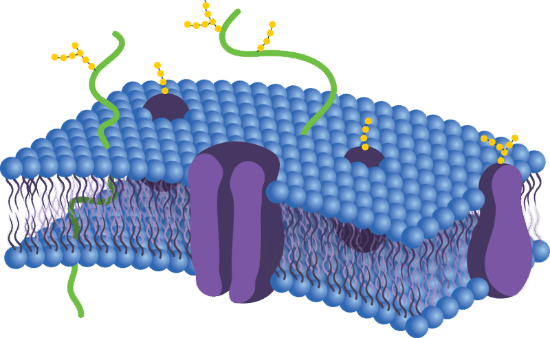
Can anything or everything move in or out of the cell?
No. It is the semipermeable plasma membrane that determines what can enter and leave the cell. So, if not everything can cross the membrane, how do certain things get across?
Membrane Proteins
The plasma membrane contains molecules other than phospholipids, primarily other lipids and proteins. The yellow molecules between the phospholipid tails in the Figure below, for example, are the lipid cholesterol. Molecules of cholesterol help the plasma membrane keep its shape. Many of the proteins in the plasma membrane assist other substances in crossing the membrane.
The plasma membranes also contain certain types of proteins. A membrane protein is a protein molecule that is attached to, or associated with, the membrane of a cell or an organelle. Membrane proteins can be put into two groups based on how the protein is associated with the membrane.
Integral membrane proteins are permanently embedded within the plasma membrane. They have a range of important functions. Such functions include channeling or transporting molecules across the membrane. Other integral proteins act as cell receptors. Integral membrane proteins can be classified according to their relationship with the bilayer:
- Transmembrane proteins span the entire plasma membrane. Transmembrane proteins are found in all types of biological membranes.
- Integral monotopic proteins are permanently attached to the membrane from only one side.
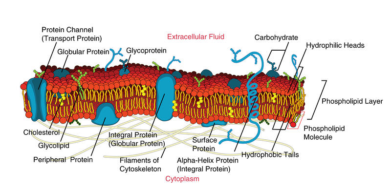
Some integral membrane proteins are responsible for cell adhesion (sticking of a cell to another cell or surface). On the outside of cell membranes and attached to some of the proteins are carbohydrate chains that act as labels that identify the cell type. Shown in the Figure below are two different types of membrane proteins and associated molecules.
Peripheral membrane proteins are proteins that are only temporarily associated with the membrane. They can be easily removed, which allows them to be involved in cell signaling. Peripheral proteins can also be attached to integral membrane proteins, or they can stick into a small portion of the lipid bilayer by themselves. Peripheral membrane proteins are often associated with ion channels and transmembrane receptors. Most peripheral membrane proteins are hydrophilic.
 Some of the membrane proteins make up a major transport system that moves molecules and ions through the polar phospholipid bilayer.
The Fluid Mosaic Model
In 1972, S.J. Singer and G.L. Nicolson proposed the now widely accepted Fluid Mosaic Model of the structure of cell membranes. The model proposes that integral membrane proteins are embedded in the phospholipid bilayer, as seen in the Figure above. Some of these proteins extend all the way through the bilayer, and some only partially across it. These membrane proteins act as transport proteins and receptors proteins.
Their model also proposed that the membrane behaves like a fluid, rather than a solid. The proteins and lipids of the membrane move around the membrane, much like buoys in water. Such movement causes a constant change in the "mosaic pattern" of the plasma membrane.
Extensions of the Plasma Membrane
The plasma membrane may have extensions, such as whip-like flagella or brush-like cilia. In single-celled organisms, like those shown in the Figure below, the membrane extensions may help the organisms move. In multicellular organisms, the extensions have other functions. For example, the cilia on human lung cells sweep foreign particles and mucus toward the mouth and nose.
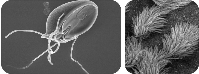 Flagella and Cilia. Cilia and flagella are extensions of the plasma membrane of many cells.
Summary
- The plasma membrane has many proteins that assist other substances in crossing the membrane.
- The Fluid Mosaic Model depicts the biological nature of the plasma membrane.
- Cilia and flagella are extensions of the plasma membrane.
Review
- What is the main difference between the two main types of proteins associated with the plasma membrane?
- What are two functions of integral membrane proteins?
- Discuss the Fluid Mosaic Model.
- What are flagella and cilia?
| Image | Reference | Attributions |
 |
[Figure 1] | Credit: Witold Ferens, based on the image Source: https://www.google.com/url?sa=i&rct=j&q=&esrc=s&source=images&cd=&ved=0ahUKEwjB95aCyanRAhVK12MKHWk8AOEQjB0IBg&url=http%3A%2F%2Fwiki.pingry.org%2Fu%2Fap-biology%2Findex.php%2FFlagella&psig=AFQjCNEEd6kumwB-KWcEschAyBt4XNpQ7g&ust=1483656311487680 License: Public Domain |
|
[Figure 2] | Credit: Mariana Ruiz Villarreal (User:LadyofHats/Wikimedia Commons) Source: commons.wikimedia.org/wiki/File:Cell_membrane_detailed_diagram_en.svg License: Public Domain |
|
[Figure 3] | Credit: Flagella: Courtesy of Dr. Stan Erlandsen/Centers for Disease Control and Prevention (Image #11643); Cilia: Courtesy of Charles Daghlian;By Mariana Ruiz Villarreal LadyofHats - the diagram i made myself using adobe illustrator. as a source for the information i used the diagrams found here:[1], [2], [3], [4], [5], [6] and [7]., Public Domain, commons.wikimedia.org/w/inde...?curid=8152684 Source: Flagella: phil.cdc.gov/phil/home.asp ; Cilia: commons.wikimedia.org/wiki/File:Bronchiolar_epithelium_3_-_SEM.jpg ; By Mariana Ruiz Villarreal LadyofHats - the diagram i made myself using adobe illustrator. as a source for the information i used the diagrams found here:[1] ; [2] ; [3] ; [4] ; [5] ; [6] and [7]. ; Public Domain ; commons.wikimedia.org/w/index.php?curid=8152684 License: Public Domain; CC BY-NC-SA 3.0 |